Chapter 21 Pelvic Pain
Pelvic pain is a frequent complaint in gynecology. It may be cyclic and associated with menstruation, sudden in onset (acute), or chronic, lasting for more than 6 months. Half of all menstruating women are affected by painful menstruation or dysmenorrhea, making it the most common type of pelvic pain. Ten percent of these women have severe symptoms necessitating time off from work or school.
Dysmenorrhea
Dysmenorrhea may be primary, when there is no readily identifiable cause, or secondary to organic pelvic disease. The typical age range of occurrence for primary dysmenorrhea is between 17 and 22 years, whereas secondary dysmenorrhea is more common in older women.
PRIMARY DYSMENORRHEA
Pathophysiology
Primary dysmenorrhea occurs during ovulatory cycles and usually appears within 6 to 12 months of the menarche. The etiology of primary dysmenorrhea has been attributed to uterine contractions with ischemia and production of prostaglandins. Women with dysmenorrhea have increased uterine activity, which results in increased resting tone, increased contractility, and increased frequency of contractions. During menstruation, prostaglandins are released as a consequence of endometrial cell lysis, with instability of lysosomes and release of enzymes that break down cell membranes.
The evidence that prostaglandins are involved in primary dysmenorrhea is convincing. Menstrual fluid from women with this disorder have higher than normal levels of prostaglandins (especially prostaglandin F2α [PGF2α] and PGE2), and these levels can be reduced to below normal with nonsteroidal antiinflammatory drugs (NSAIDs), which are effective treatments. Infusions of PGF2α or PGE2 reproduce the discomfort and many of the associated symptoms such as nausea, vomiting, and headache. Secretory endometrium contains much more prostaglandin than proliferative endometrium. Women with primary dysmenorrhea have upregulated cyclooxygenase (COX) enzyme activity as a major cause of their pain. Anovulatory endometrium (without progesterone) contains little prostaglandin, and these menses are usually painless.
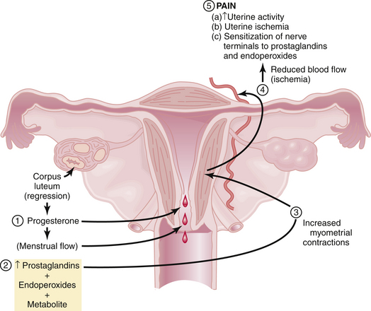
Figure 21-1 summarizes the relationships among endometrial cell wall breakdown, prostaglandin synthesis, uterine contractions, ischemia, and pain.

FIGURE 21-1 Postulated mechanism of pain generation in primary dysmenorrhea. Nonsteroidal anti-inflammatory drugs inhibit cyclooxygenase, the enzyme that catalyzes the formation of prostaglandins from arachidonic acid. Hormonal contraceptives that block ovulation significantly reduce the formation of prostaglandins. Both drugs mitigate this mechanism of pain and are effective treatment for primary dysmenorrhea.
(Modified from Dawood MY: Hormones, prostaglandins and dysmenorrhea. In Dawood MY [ed]: Dysmenorrhea. Baltimore, Williams & Wilkins, 1981.)
Clinical Features
The clinical features of primary dysmenorrhea are summarized in Box 21-1. Cramping usually begins a few hours before the onset of bleeding and may persist for hours or days. It is localized to the lower abdomen and may radiate to the thighs and lower back. The pain may be associated with altered bowel habits, nausea, fatigue, dizziness, and headache.
Treatment
Box 21-2 lists the treatment options for primary dysmenorrhea. NSAIDs, which act as COX inhibitors, are highly effective in the treatment of primary dysmenorrhea. Typical examples include ibuprofen (400 mg every 6 hours), naproxen sodium (250 mg every 6 hours), and mefenamic acid (500 mg every 8 hours). Decreasing prostaglandin production by enzyme inhibition is the basis of all NSAIDs. Hormonal contraceptives such as oral contraceptive pills (OCs), patches, or transvaginal rings reduce menstrual flow and inhibit ovulation and are also effective therapy for primary dysmenorrhea. Extended cycle use of OCs or the use of long-acting injectable or implantable hormonal contraceptives or progestin-containing intrauterine devices minimizes the number of withdrawal bleeding episodes that users have. Some patients may benefit from using both hormonal contraception and NSAIDs.
Resistant cases may respond to tocolytic agents (e.g., salbutamol), a calcium blocker (e.g., nifedipine), or high-dose continuous daily progestogens (especially medroxyprogesterone acetate or dydrogesterone). Nonpharmacologic pain management, particularly acupuncture or transcutaneous electrical stimulation, may be useful, as are psychotherapy, hypnotherapy, and heat patches. Surgical procedures such as presacral neurectomy and uterosacral ligament section have been largely abandoned.
If a patient fails to respond to hormonal contraception and NSAID therapy, the diagnosis of primary dysmenorrhea should be questioned and consideration given to a secondary cause. Ultrasonic imaging, laparoscopy, and hysteroscopy with directed biopsy should be performed to exclude pelvic disease.
SECONDARY DYSMENORRHEA
Pathophysiology
The mechanism of pain in secondary dysmenorrhea depends on the underlying (secondary) cause and in most cases is not well understood. Prostaglandins may also be involved in this type of dysmenorrhea, although NSAIDs and hormonal contraceptives that do not suppress menses altogether are less likely to provide satisfactory pain relief.
Clinical Features
The clinical features of some of the underlying causes of secondary dysmenorrhea are summarized in Box 21-3. In general, secondary dysmenorrhea is not limited to the menses and can occur before as well as after the menses. In addition, secondary dysmenorrhea is less related to the first day of flow, develops in older women (in their 30s or 40s), and is usually associated with other symptoms such as dyspareunia, infertility, or abnormal uterine bleeding.
Treatment
Management consists of the treatment of the underlying disease. The treatments used for primary dysmenorrhea (Box 21-2) are often helpful. Other specific treatments are discussed in the chapters dealing with the underlying causes.
Acute Pelvic Pain
Acute pain is sudden in onset and is usually associated with significant neuroautonomic reflexes such as nausea and vomiting, diaphoresis, and apprehension. It is important for the gynecologist to be aware of both the gynecologic and nongynecologic causes of acute pelvic pain (Box 21-4). Delay of diagnosis and treatment of acute pelvic pain increase the morbidity and even mortality.
Adnexal accidents, including torsion or rupture of an ovarian (Figure 21-2) or fallopian tube cyst, can cause severe lower abdominal pain. Normal ovaries and fallopian tubes rarely undergo torsion, but cystic or inflammatory enlargement predisposes to these adnexal accidents. The pain of adnexal torsion can be intermittent or constant, is often associated with nausea, and has been described as reverse renal colic because it originates in the pelvis and radiates to the loin. An enlarging pelvic mass is found on examination and ultrasound with decreased or absent blood flow to the adnexa on Doppler ultrasound studies. The need for surgical intervention is common and urgent.
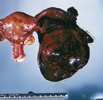
FIGURE 21-2 Torsion of an ovarian cyst.
(From Clement PB, Young RH: Atlas of Gynecologic Surgical Pathology. Philadelphia, WB Saunders, 2000.)
Functional ovarian cysts (e.g., corpus luteum or follicular cysts) may rupture, causing leakage of fluid or blood that causes acute pain from peritoneal irritation. When there is significant associated bleeding, the pain may be followed by a hemoperitoneum and hypovolemia. Surgical intervention is mandatory in this setting, after adequate resuscitation with packed red cells and intravenous fluids.
Acute reproductive organ infections such as endometritis

Stay updated, free articles. Join our Telegram channel

Full access? Get Clinical Tree