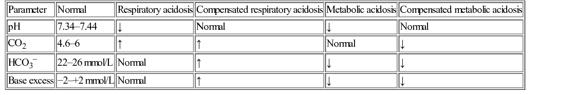
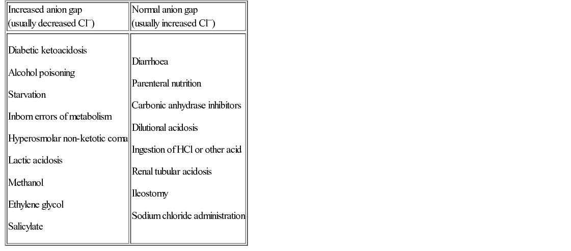
Lynn Sinitsky, Michael Marsh, David Inwald • Understand the science underpinning basic and advanced life support • Know how to interpret blood gas abnormalities • Understand the pathophysiology of respiratory failure • Understand the scientific basis of respiratory support including mechanical ventilation in children • Know about the basis for the recognition of dehydration and fluid management • Know how to interpret fluid and electrolyte abnormalities • Understand the pathophysiology of shock and its management • Understand the pathophysiology of traumatic and other encephalopathies and their management The outcome of cardiac arrest in children is poor. To minimize the incidence of cardiac arrest and its consequent morbidity and mortality, a structured approach to children with serious illness or injury is required (Box 6.1). This also aids communication between health professionals and provides a safe and effective methodology for assessment of the seriously unwell child. The primary assessment systematically looks at respiratory, cardiovascular and neurological status and addresses problems as they are found. A secondary assessment is then performed, looking at each system in turn and instituting emergency treatment. This approach, pioneered by the Advanced Life Support Group (ALSG) has been shown to be effective in reducing mortality. Following an ALSG education programme in Gambia, there was a 50% reduction in maternal mortality, a 32% reduction in infant mortality and a 94% survival rate in resuscitation. As part of recognition and management of critically ill children, whatever the underlying cause, the following physiological variables should be monitored: They provide important additional information to the clinical observations of ABCD. They need to be related to normal age-appropriate values (see Chapter 3, History and examination). They also help to answer the important ongoing questions: • Are things getting better or worse? • If worse, do I need to escalate treatment and get senior involvement? Although paediatric life support courses have been shown to improve health professionals’ ability to recognize and provide initial management of critically ill children, regular reinforcement is required to maintain these skills. There are many anatomical airway differences between infants, children and adults (see Box 6.2 and also Chapter 17, Respiratory medicine). The younger the child, the more pronounced the difference. This has relevance to emergency care, in particular airway opening manoeuvres, airway adjuncts and endotracheal intubation. Life-threatening airway obstruction can develop rapidly in children as the airway is already narrow and further airway narrowing from any cause increases resistance according to Pouseuille’s law (see Chapter 17, Respiratory medicine, for a detailed discussion). As resistance is inversely proportional to the fourth power of the radius, small reduction in their already small airway radius will result in a large increase in resistance. Due to the relatively large head and short neck in infants, neck flexion or overextension can cause airway obstruction by tracheal compression. The relatively large tongue can also cause airway obstruction, especially if there is a reduced level of consciousness, as well as impede the view at laryngoscopy. Airway opening manoeuvres, the head tilt/chin lift and jaw thrust manoeuvres, are used to improve patency of an obstructed or partially obstructed airway. Both manoeuvres apply anterior tension to the hyoid bone and draw the epiglottis away from the posterior pharyngeal wall, opening the pharynx. In addition, the jaw thrust manoeuvre pulls the tongue, which may cause airway obstruction, away from the palate and opens the oropharynx. Care must also be taken when positioning fingers for airway opening manoeuvres as the floor of the infant’s mouth is easily compressible. In the head tilt/chin lift manoeuvre, it is important to control the degree of head tilt to avoid airway narrowing due to overextension of the head and neck. In the infant, due to their large head and short neck, a neutral position is preferred. In the child, the sniffing position is used. This manoeuvre is contraindicated if there is history of trauma because it may exacerbate cervical spine injury. Therefore only jaw thrust is recommended in the airway management of paediatric trauma. An oropharyngeal airway adjunct or Guedel airway creates an open channel between the mouth to the posterior pharyngeal wall. They are only used in infants and children with a reduced level of consciousness as they may cause choking or vomiting if the gag reflex is present. They are sized according to length, by measuring the distance between the anterior nares and tragus of the ear. Airway adjuncts that are too small may be ineffective and those too large may cause laryngospasm. A nasopharyngeal airway adjunct is often better tolerated than an oropharyngeal airway. However, insertion may cause haemorrhage from the vascular nasal mucosa and worsen airway obstruction. It is contraindicated in basal skull fracture. A laryngeal mask airway is a device for supporting and maintaining the airway without tracheal intubation (Fig. 6.1). It is designed to sit in the hypopharynx and cover the supraglottic structures, thereby allowing relative isolation of the trachea. In the emergency setting, it is helpful for airway obstruction caused by supraglottic airway abnormalities or if bag-mask ventilation is not possible. A laryngeal mark does not totally protect the airway from aspiration of secretions, blood or stomach contents, and with high pressure ventilation, gastric distension may occur. Endotracheal intubation can be difficult in infants and should only be performed by trained health professionals. Unless the child is unresponsive, it should be preceded by induction of anaesthesia using drugs for sedation and neuromuscular blockade. There may be clues in the history or examination that suggest a child has a difficult airway (Box 6.3). Under these circumstances, senior anaesthetist and ENT surgeon should be present. An understanding of the anatomy and appropriate preparation of equipment aids success. In infants, the epiglottis is horse-shoe shaped and relatively large. In addition, the larynx is high anterior (at C2/C3 in infants compared with C5/C6 in older children/adults). A straight blade laryngoscope (‘Miller blade’) is therefore more commonly used in infants, positioned posterior to the epiglottis, lifting it to allow visualization of the glottis and vocal folds (the ‘Miller lift’) (Fig. 6.2A). A curved blade laryngoscope (‘Macintosh’) is used in children and adults, and is positioned in the vallecula, anterior to the epiglottis, lifting it to visualize the larynx (Fig. 6.2B). Endotracheal tubes are sized by internal diameter in mm. Until the age of 12 years, the narrowest part of the airway is at the level of the cricoid cartilage. Therefore, an endotracheal tube that passes easily through the vocal cords may still be too large to pass through the cricoid ring. Too tight a fit at this level may damage the mucosa. Subsequent airway oedema and post-extubation stridor can occur and, rarely, tracheal scarring and stenosis may follow. For many years, the use of cuffed tubes in children under 8 years old was considered inappropriate because of fears of mucosal damage at the level of the cricoid cartilage. However, over the last 10 years, studies have demonstrated that cuffed endotracheal tubes can be used safely in infants and young children providing correct tube size is used, tube position verified and cuff inflation pressure checked and limited. Consequently, cuffed endotracheal tubes are now first choice in paediatric critical care and are preferred in children with poor lung compliance or high airway resistance, or when precise ventilation and/or CO2 control is needed. Uncuffed tubes are still used in newborn infants. Cardiorespiratory arrest in children is rare (incidence 1–20 per 100,000 children per year in developed countries). The majority occur in children under the age of one year. In adults, cardiac arrest is often due to primary cardiac disease, which occurs with near-normal function of the circulatory and respiratory systems until the moment of arrest. In contrast, in children, most occur secondary to hypoxia due to respiratory failure or circulatory failure due to fluid loss or fluid maldistribution. End organ damage is therefore often already present in children at the time of cardiac arrest and is responsible for their prognosis. However, in up to 30% of children, there is a cardiac cause for cardiopulmonary arrest. This is becoming more common with improved survival in children with complex congenital heart disease. There are also children with sudden unexpected ventricular fibrillation (VF) or pulseless ventricular tachycardia (VT) who may have previously unrecognized cardiac disease, either a cardiomyopathy or an inherited channelopathy. Following an unexpected cardiac arrest or VF/VT arrest, referral to a paediatric cardiologist should be made. An identifiable cause may be found following detailed investigation, which may include genetic studies and pharmacological provocation tests, and treatment given. As family members of these children may also be at risk of sudden death, they should also be referred. Cardiopulmonary resuscitation (CPR) is an emergency procedure to delay cell damage and death in the heart and brain by facilitating partial flow of oxygenated blood to these organs. This provides a brief window of opportunity to restore breathing and spontaneous blood circulation. CPR comprises chest compressions and breaths, applied in a ratio of 15 compressions to 2 breaths (except in newborn infants, for whom a 3 : 1 ratio is required). Adequate chest compressions should be provided at a depth and rate that allow complete recoil of the chest after each compression. Experimental and mathematical studies have shown that the best compression rate at all ages is 100–120 per minute. The recommended depth is one third of the depth of the chest. There should be minimal interruption of compressions as coronary perfusion pressure has been shown to be greater with prolonged continuous compressions. In adults, survival rates from out-of-hospital cardiorespiratory arrests are improved if bystander CPR is given, even if it is compression-only CPR. In children, bystander CPR also results in a more favourable outcome, but not if it is compression-only CPR. As most out-of-hospital arrests in children are hypoxic in origin, rescue ventilation remains the cornerstone of paediatric CPR. Prolonged resuscitation is, in general, associated with a bad neurological outcome. However, a recent study of successful CPR in adults has shown neurological outcome is not directly correlated to the duration of CPR. Following this, the Resuscitation Council (UK) does not recommend a specific duration for CPR. Instead, clinicians are to determine duration on a case-by-case basis, continuing prolonged resuscitation where there is potential for a reversible cause. Unfortunately, studies in children have not reported similar favourable outcomes in prolonged resuscitation, unless there is associated profound hypothermia (<30°C) or intermittent return of spontaneous circulation (ROSC). Survival is rare if resuscitation continues for longer than 20–30 minutes and any survivors are likely to have significant neurological deficits. However, there are cases of survival with reasonable outcome following prolonged in-hospital cardiac arrest in cardiac centres with ECMO (extra-corporeal membrane oxygenation). Although such specialist treatment is not feasible for all children following cardiopulmonary arrest, it raises an interesting question about the potential for treatments other than conventional CPR in the management of cardiopulmonary arrest. Following return of circulation, children who survive cardiorespiratory arrest have significant multi-organ dysfunction. They should be transferred to an intensive care setting for specialist post-resuscitation care. Management focuses on achieving and maintaining homeostasis in order to optimize multi-organ recovery; initiating investigation of the underlying cause of the respiratory arrest; and treating any identifiable cause. There is increasing evidence that hyperoxaemia can be detrimental. Excessive tissue oxygen concentrations may increase production of oxygen free radicals. Oxygen free radicals can damage mitochondria and so there is potential to compound neuronal damage. During resuscitation beyond the neonatal period, 100% oxygen is used. However, after return of spontaneous circulation, inspired oxygen should be titrated to achieve oxygen saturations of 94–98% using pulse oximetry. In adults, mild therapeutic hypothermia has been shown to improve neurological outcome after ventricular fibrillation (VF) arrest. Therapeutic hypothermia of newborns with hypoxic–ischaemic encephalopathy has been shown to be associated with improved neurological outcome. Although the role of therapeutic hypothermia post cardiac arrest in children remains unclear, current guidelines suggest therapeutic cooling to 32–36°C for at least 24 hours. As increased core temperature increases metabolic demand by 10–13% for each degree centigrade above normal, as a minimum, hyperthermia should be avoided or, if present, treated with active cooling to achieve a normal core temperature. Shivering should be avoided as it increases metabolic demand. Sedation and neuromuscular blockade may be needed. In all age groups, abnormal blood glucose levels (hyper- or hypoglycaemia) are associated with poor outcome following cardiorespiratory arrest. It remains unclear whether this is a causative or associated factor. Following the return of spontaneous circulation, plasma glucose levels should be monitored and hypo- and/or hyperglycaemia avoided. However, tight glucose control is not recommended as this increases the risk of hypoglycaemia without any survival benefit. For children who have a respiratory arrest without cessation of circulation, whether out-of-hospital or in-hospital, more than two thirds survive. The majority who get to paediatric intensive care (PICU) survive, most (over 90%) with a favourable neurological outcome. However, outcome from cardiopulmonary arrest is poor. Only a third get to PICU. Of these, only a third survive to discharge from hospital, the majority (up to 90%) with moderate to severe neurological deficit due to hypoxic–ischaemic brain injury. Discussion of withdrawal or limitation of life-sustaining treatment may be difficult and emotive. All health professionals have a duty to act in the best interests of the child. When treatment neither restores health nor confers any other benefit it may no longer be in the child’s best interests. Sympathetic discussion between health professionals and parents usually allows a joint decision to evolve. Guidance is available from the General Medical Council (GMC), British Medical Association (BMA) and Royal College of Paediatrics and Child Health (RCPCH), which also addresses the uncommon situation when a joint decision cannot be reached. This is considered in more detail in Chapter 35, Ethics. Acid base disorders are very common in critically ill and injured children. A brief reminder of the principles is outlined below, and Table 6.1 summarizes common arterial blood gas patterns. pH is defined as the decimal logarithm of the reciprocal of the hydrogen ion activity or concentration (aH+) in a solution: pH is maintained in range (7.34–7.44) by several buffering systems, the main one being the carbonic acid–bicarbonate system: The relationship between pH, HCO3 and CO2 is described by the Henderson–Hasselbalch equation: In hyperventilation, CO2 is blown off and pH increases (respiratory alkalosis). In hypoventilation, CO2 is retained and the blood becomes acidotic (respiratory acidosis). Respiratory changes have a rapid effect on the pH. HCO3 is a base and a buffer of hydrogen ions (H+), which are the product of normal cellular metabolism. When an excess of hydrogen ions are present due to excess acid production, the buffering effect of HCO3 is overcome and the blood becomes acid (metabolic acidosis). Examples of excess acid are lactic acid in shock or ketones in diabetic ketoacidosis, acid administration (e.g. salicylic acid in aspirin poisoning) or bicarbonate loss (e.g. from the gut in gastroenteritis or in the urine in renal tubular disease). The kidneys can compensate for a respiratory acidosis in chronic respiratory failure by increasing the amount of HCO3 in the blood and extracellular fluid (compensated respiratory acidosis). This results in a normal pH and a high CO2 and HCO3. This compensation takes several days. The respiratory system can compensate for a metabolic acidosis by blowing off CO2 to normalize pH (compensated metabolic acidosis). This response is mediated via carotid chemoreceptors and results in normal pH, low CO2 and low HCO3. The main limitation of the Henderson–Hasselbalch approach is that buffers other than HCO3 exist, including haemoglobin and albumin. The consequence of this is that HCO3 and CO2 are not independent. In the dissociation equation below, a rise in CO2 will cause formation of hydrogen ions: However, as hydrogen ions are buffered by haemoglobin and albumin, bicarbonate levels also rise. Thus, a rise in CO2 results in a rise in HCO3. This rise in HCO3 could be mistaken for a metabolic alkalosis, when the cause was respiratory acidosis. The concept of base excess was developed to address this problem. Base excess is determined by equilibrating the sample to a normal pCO2 (5.33 kPa) then titrating it to pH 7.4. The number of mmol/L required to do this equals the base excess, and is therefore a measure of how acidotic or alkalotic the sample is without any contribution of CO2. Finally, calculation of the anion gap (= [Na+] + [K+]) − ([Cl−] + [HCO3−]) allows classification of a metabolic acidosis into those with a normal or increased anion gap. The anion gap is a measure of the concentration of unmeasured anions (e.g. plasma proteins, ketones, lactate) and is based on the theory of electrical neutrality (the sum of the positive ions must equal the sum of the negative ions). An increased anion gap (>16 mmol/L) suggests the presence of unmeasured organic acid, whereas a normal anion gap implies bicarbonate loss and/or increase in chloride concentration (Table 6.2; see Fig. 29.1). Hyperchloraemic acidosis is common in patients given normal saline-containing fluids. There are two reasons for this. Firstly, normal saline (pH 5–6) is acidic and has little buffering capacity. The acidic pH of normal saline is due to the ‘Grotthuss mechanism’, whereby ions which dissociate when dissolved in water (e.g. Na+ and Cl− ions) cause disruption of the ionic bonding of H2O, leading to greater dissociation and generation of [H+]. Secondly, volume expansion causes plasma bicarbonate dilution. The importance of recognizing hyperchloraemia as a cause of acidosis is to avoid administration of further normal saline-containing fluids when further fluid therapy is unnecessary and will exacerbate the problem. The finding of a normal anion gap, raised chloride concentration and low bicarbonate should be the clue.
Paediatric emergencies and critical care
Recognition and management of the seriously ill or injured child
Monitoring and post-resuscitation management
The science of basic and advanced life support
The paediatric airway
Airway opening manoeuvres
Use of airway adjuncts
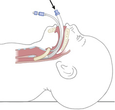
Laryngeal mask airway
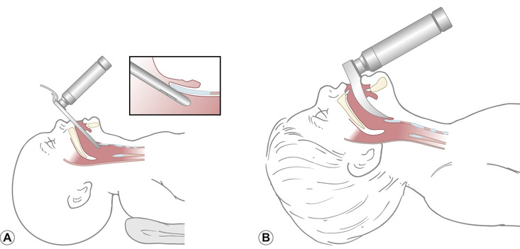
Endotracheal intubation
Cardiopulmonary arrest
Causes of cardiopulmonary arrest
Treatment of cardiopulmonary arrest
Cardiac compressions and ventilation breaths
Duration of resuscitation
Post-resuscitation care
Oxygen
Therapeutic hypothermia
Blood glucose control
Resuscitation outcomes
Blood gas abnormalities


Hyperchloraemic acidosis
Scientific basis of respiratory support including mechanical ventilation
What is respiratory failure?
Respiratory failure may be defined as a syndrome of inadequate gas exchange, with the result that levels of arterial oxygen, carbon dioxide or both cannot be maintained within their normal ranges (Box 6.4). A drop in arterial oxygenation is hypoxaemia; a rise in arterial carbon dioxide levels is hypercapnia. Classification into type I or type II relates to the absence or presence of hypercapnia, respectively.
Type 1 respiratory failure is defined as hypoxia without hypercapnia, and the PaCO2
Stay updated, free articles. Join our Telegram channel

Full access? Get Clinical Tree


