Fig. 13.1
Transvaginal ultrasound of the left ovary revealed an intraovarian gestational sac with yolk sac adjacent to suspected corpus luteum. (Reproduced with permission from The Journal of Minimally Invasive Gynecology)

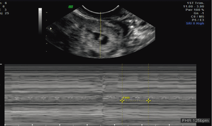
Fig. 13.2
Transvaginal ultrasound of the left ovary revealed an intraovarian gestational sac with detectable fetal heart rate with M-mode. (Reproduced with permission from The Journal of Minimally Invasive Gynecology)
My Management
a.
Administer single-dose methotrexate 50 mg intramuscularly and check hCG levels on days 4 and 7 to ensure downward trend.
b.
Perform laparoscopy and left (salpingo) oophorectomy to optimize treatment and confirm diagnosis of ovarian pregnancy histopathologically .
c.
Administer methotrexate locally directly into the pregnancy via transvaginal ultrasound guidance.
Diagnosis/Assessment
Implantation of a pregnancy outside of the endometrium, collectively referred to as an ectopic pregnancy, complicates approximately 2 % of pregnancies [2]. Nontubal ectopics are especially concerning since they may present late and/or be more vascularized with a higher risk of life threatening bleeding if they rupture. Primary ovarian ectopic pregnancy (OEP) is a rare form of ectopic pregnancy, estimated to occur in 3.6 % of all ectopic pregnancies [3]. The etiology of primary OEP remains unknown and can occur in the absence of risk factors. Embryo migration from the fallopian tube has been described as a potential mechanism of an ovarian ectopic [4]. A review by Joseph and Irvine reported that contraceptive intrauterine devices and assisted reproductive technology each accounted for roughly 20 % of all ovarian ectopic pregnancies [5].
Since many women with ectopic pregnancies are asymptomatic, early diagnosis of ectopic pregnancy, especially nontubal ectopic pregnancies, requires a high index of suspicion and a skilled ultrasonographer. Abdominal pain and vaginal bleeding are presenting symptoms of an ectopic pregnancy regardless of the location, making it difficult to distinguish an OEP from both tubal and nontubal pregnancies. Failure to diagnose an OEP can be catastrophic due to potential rupture of the ovary and subsequent internal bleeding , dramatically increasing the likelihood of needing major surgical intervention alongside loss of the ovary. Indeed, ruptured ectopics may present with an acute abdomen, shoulder pain (secondary to diaphragmatic irritation), and even hypovolemic shock secondary to the hemoperitoneum [5].
The classic diagnosis of an OEP was confirmed surgically by the original four Spiegelberg criteria (1878): (1) the ipsilateral fallopian tube and fimbria are intact and separate from the ovary, (2) the gestational sac is in the normal position of the ovary, (3) the gestational sac is connected to the uterus by the ovarian ligament, and (4) ovarian tissue must be attached to the specimen and within the gestational sac. Although the classic definitive diagnosis of an OEP depends on these histopathologic findings obtained surgically [6], advances in transvaginal sonography have led it to become a key diagnostic tool in ovarian, as well as other, ectopic pregnancies. Ovarian pregnancies classically appear as a hypoechoic cyst on or within the ovary and are characterized by a wide echogenic (hyperechoic) outside ring [7]. A yolk sac or fetal pole is less commonly seen. Interestingly, approximately 70 % of ovarian ectopic pregnancies are not diagnosed, instead being mistaken as a ruptured corpus luteum or a hemorrhagic cyst due to their similar clinical features [6]. In the case presented here, OEP was diagnosed by sonographic evidence of a gestational sac and fetal pole with a measurable heart rate within the left ovary.
Management
Management strategies used for OEPs are similar to those used in tubal pregnancies [5]. Hemodynamic stabilization with immediate surgical management is appropriate with clinical features suggestive of a ruptured ectopic pregnancy in the unstable patient. The surgical approach is both patient- and surgeon-dependent, consisting of laparoscopy and/or laparotomy with the ultimate goals of removal of ectopic pregnancy tissue, achieving hemostasis , and preservation of healthy ovarian tissue [8]. Historically, OEPs were treated via removal of the entire ovary or by wedge resection attempting to conserve some remaining healthy ovarian tissue [9]. Postoperative methotrexate may be considered with concern for residual trophoblastic tissue following surgery in appropriate patients [5].
Stay updated, free articles. Join our Telegram channel

Full access? Get Clinical Tree


