Chapter 68 Orthopedics
A Practical Approach
NEWBORN INFANT
ETIOLOGY
What Common Orthopedic Problems Affect Newborns?
Almost all newborns have bowing or internal curvature of the legs to some degree. Crossing one’s arms and placing each hand on the opposite shoulder simulates the intrauterine position of the legs and feet of the fetus, readily explaining this transient deformity seen in the newborn. There is no force to correct this bowing until the child begins to bear weight on the legs for standing and walking. Parental reassurance may be needed.
What Is Developmental Dysplasia of the Hip?
Developmental dysplasia of the hip (DDH) is the orthopedic condition most likely to cause an adverse outcome unless diagnosed in early infancy. Identification of DDH is one of the most important goals of the newborn examination. Formerly called congenital dislocation of the hip, DDH refers to dislocation of the femoral head from the acetabulum because of incomplete formation of the acetabular roof. Predisposing factors include having a family history of DDH, being first-born, being female (9:1 female/male ratio), or being in the breech position in utero.
What Foot Problems Are Identified in the Newborn?
Metatarsus varus is a common finding, whereas talipes equinovarus (clubfoot) occurs uncommonly. Metatarsus varus, also referred to as metatarsus adductus or “C-foot,” most often is a flexible positional deformity and less commonly is a rigid structural deformity. Clubfoot is a rigid deformity of the bony structures in the ankle and foot. It can be idiopathic (75%) or may occur in association with other neuromuscular disorders.
EVALUATION
How Do I Detect Developmental Dysplasia of the Hip?
Careful examination of the hips is mandatory for all newborns. The Ortolani maneuver identifies the dislocated hip, and the Barlow maneuver identifies the dislocatable hip. Careful attention to proper technique is critical (See Chapter 5 and Figure 5-4). You must be able to distinguish DDH from a capsular or ligamentous click, which is a benign finding similar to the “popping” that might occur in your own joints. This click is very different from the gross “clunk” felt when a hip is reduced or dislocated by the Ortolani or Barlow maneuver, respectively. The examination for DDH is done on each physical examination until the age of 1 year, but after age 2 to 3 months, the dislocated hip cannot be reduced by the Ortolani maneuver, and limited abduction is the abnormal finding in DDH. Ultrasonography is the preferred imaging modality when DDH is suspected.
How Do I Distinguish the Various Foot Deformities?
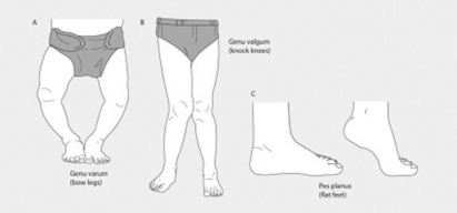
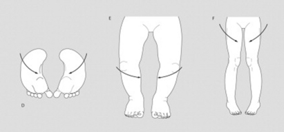
All foot deformities are identified by visual inspection and by manipulation to determine whether the deformity is flexible or rigid. The lateral border of the sole of the foot should be a straight line. Metatarsus varus (Figure 68-1D) is present if the foot curves inward (like the letter C). If the curve can be easily straightened, it represents a flexible deformity. A fixed deformity cannot be easily straightened. Talipes equinovarus or clubfoot requires three features for diagnosis: hindfoot equinus (plantarflexion of the foot at the ankle), hindfoot varus (inversion deformity of the heel), and forefoot varus. This deformity cannot be straightened by simple manipulation.
TREATMENT
Does Developmental Dysplasia of the Hip Require Surgical Correction?
If DDH is diagnosed early, stabilization of the femoral head in the acetabulum by use of a harness that maintains the femurs in abduction is curative. The Pavlik harness is one example of such a treatment device. Use of “double” or “triple” diapers is not an acceptable treatment for the newborn with DDH. In more severe cases, generally meaning those in which recognition was delayed, DDH is managed by casting or surgery, and the likelihood of a good outcome is less.
Is Treatment Needed for Physiologic Deformities?
Physiologic or positional deformities of the foot and tibia do not require any treatment. Flexible metatarsus varus is often “treated” by daily stretching to straighten the lateral border of the foot, but there is little evidence that such manipulation hastens correction of the curve. Advise patience and remind parents that tibial bowing will correct when the child begins to bear weight on the legs for standing and walking.
How Are Rigid Deformities Managed?
Rigid metatarsus varus will respond to casting for 2 to 3 weeks. This is generally best left to an orthopedist familiar with pediatric problems. Talipes equinovarus is a much more serious problem that can significantly impair ankle and foot function if left untreated. Current management involves serial casting and long-term orthopedic follow-up. Surgical repair may be needed in the most severe cases.
OLDER INFANTS AND TODDLERS
ETIOLOGY
What Orthopedic Problems Affect This Age Group?
Older infants and toddlers commonly have deformities of the extremities, mainly of the legs, that provoke parental concern. Most do not cause long-lasting problems and do not require any treatment, but you must be able to identify them so that you can reassure worried parents. The deformities reflect physiologic variations of “normal” and include either angulation at a joint or rotation along a long bone (Figure 68-1). Angular deformities affect the knees (genu valgus and genu varus) and the feet (metatarsus varus—also called metatarsus adductus). Although metatarsus varus should be identified and managed in the newborn, it may be undetected until the child begins to walk and parents experience problems finding shoes that fit. Rotational deformities affect the femur (femoral anteversion or retroversion) and the tibia (tibial torsion). Femoral anteversion describes internal rotation of the femur, and femoral retroversion describes external rotation. Both cause the feet to turn away from the expected straight position and may be associated with tripping or gait patterns that concern parents. Internal tibial torsion (ITT) refers to rotation along the long axis of the bone, not bowing. The rotation of ITT is toward the midline, resulting in the feet also “turning in” toward the midline.
What Is Nursemaid’s Elbow?
The term nursemaid’s elbow refers to subluxation of the head of the radius. This common toddler injury often happens when the toddler trips while holding an adult’s hand. The adult reflexively attempts to bring the falling child back to his or her feet. The child’s arm is sharply pulled against resistance, which causes the annular ligament to slide over the head of the radius. The child dramatically stops using the arm and complains of pain.
EVALUATION
How Are Positional Leg Deformities Detected?
Parents often bring these deformities to the attention of the physician by expressing concern that “toes turn in” or that “the legs are bowed.” “Tripping” is another concern brought up by parents of toddlers. Whether angular or rotational, positional deformities are detected by inspection of the affected extremity and comparison with the opposite. Knowledge of the range of physiologic variation is important and is described for the specific conditions that follow. Watching the child walk helps to identify the problem.
How Do I Describe an Angular Deformity?
The first term of a condition names the anatomic location of the extremity, such as metatarsus; the second term describes the position of the extremity distal to that location in relationship to the midline. Varus means tilted toward the midline. Valgus means tilted away from the midline. Therefore, the term metatarsus varus indicates that the part of the foot distal to the metatarsal is tilted toward the midline. Applying this terminology scheme, does the term genu valgus describe knock-knee or bowleg?
How Do I Detect Internal Tibial Torsion?
ITT is most objectively assessed by palpation of the tibial tuberosity in the midline of the tibia to center the leg. Then palpate the medial and lateral malleoli. The medial malleolus should be 10 to 15 degrees anterior to the lateral malleolus in the horizontal plane. In the presence of moderate ITT, the malleoli are in the same plane. With severe internal torsion, the lateral malleolus is anterior. When affected children are viewed straight-on while standing, the knees seem to be aligned, but the feet are turned inward.
How Do I Detect Femoral Rotational Deformities?
The range of rotation of the femur is 100 to 110 degrees. With the child lying supine, grasp each lower leg, then internally and externally rotate the femurs to determine if rotation is excessive in either direction. Very few children (or adults) have equal inward and outward rotation of the femur. Most individuals have some degree of either excessive internal or external rotation. In contrast to ITT, when affected children are viewed straight on while standing, both the knees and the feet turn inward (or outward).
What Will I Find on Examination of a Child with Nursemaid’s Elbow?
The child usually refuses to use the arm, holding it in a pronated and slightly flexed position, and resists any movement. The key to recognition is the history of sudden pulling or jerking of the arm. Because of lack of ossification of the radial head, no radiographic abnormality is visible, so radiologic studies are not needed.

Stay updated, free articles. Join our Telegram channel

Full access? Get Clinical Tree

