OPHTHALMOLOGY

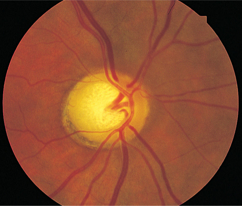
Figure 14-1. Optic disk cupping in glaucoma. (Reproduced, with permission, from Longo DL, Fauci AS, Kasper DL, et al, eds. Harrison’s Principles of Internal Medicine. 18th ed. New York: McGraw-Hill; 2012).
What is the most likely diagnosis?
a. Open-angle glaucoma
b. Idiopathic intracranial hypertension (IIH)
c. Angle-closure glaucoma
d. Diabetic retinopathy
e. Optic nerve drusen
Answer a. Open-angle glaucoma
Open-angle glaucoma is a disorder that results in increased pressure in the eye. Open angle chronic glaucoma is painless and develops over time. On examination there is optic disc cupping describes a hollowed-out appearance of the optic nerve or “disc” on funduscopy. A cup whose diameter is greater than 50% of the vertical disc diameter is indicative of glaucoma. IIH has optic disc blurring; angle-closure glaucoma is painful and presents with a fixed red eye. Diabetic retinopathy has neovascularization and cotton wool spots, and optic nerve drusen has globules of mucoproteins and mucopolysaccharides that progressively calcify in the optic disc.
Aqueous humor → trabecular meshwork → canal of Schlemm
The U.S. Preventive Services Task Force does not recommend screening for glaucoma.
What is the best next step to confirm the diagnosis in of this patient?
a. Tonometry
b. Visual field defects
c. Tonometry and visual field defects
Answer c. Tonometry and visual field defects
Tonometry alone is not enough to diagnose glaucoma, and neither is visual field defects alone; however, together their sensitivity and specificity are high enough to confirm glaucoma. Furthermore, the absence of nerve damage on funduscopy does not rule out open-angle glaucoma because many patients with elevated pressure do not have evidence of nerve damage. For cases in which patients are equivocal on both, corneal thickness measurement or pachymetry is the tiebreaker.
Intraocular pressure (IOP) and what to do!
• IOP >40 mm Hg: emergency referral
• IOP 30 to 40 mm Hg: urgent referral to ophthalmology office
• IOP 25 to 29 mm Hg: evaluation within 1 week
• IOP 23-24 mm Hg: repeat measurement in 6 months
What is the best next step in the management of this patient?
a. Pharmacologic therapy
b. Laser therapy
c. Surgical therapy
Answer a. Pharmacologic therapy
Lowering IOP has been shown to reduce the risk of progression of visual field loss and optic disc changes. There are two major kinds of medications: the first increases aqueous outflow, and the second decreases aqueous production. Laser therapy is second-line treatment and increases aqueous outflow. Surgical therapy is for refractory cases and creates a filtration bleb to allow aqueous humor to leave the eye through an alternative route.
Prostaglandin analogs, such as latanoprost, increase outflow of aqueous humor.
β-Blockers, such as timolol, decrease aqueous humor production.
Carbonic anhydrase inhibitors, such as acetazolamide, inhibit carbonic anhydrase in the ciliary body and decrease aqueous humor production.
What is the most likely diagnosis?
a. Conjunctivitis
b. Corneal abrasion
c. Infectious keratitis
d. Angle-closure glaucoma
e. Traumatic hyphema
Answer d. Angle-closure glaucoma
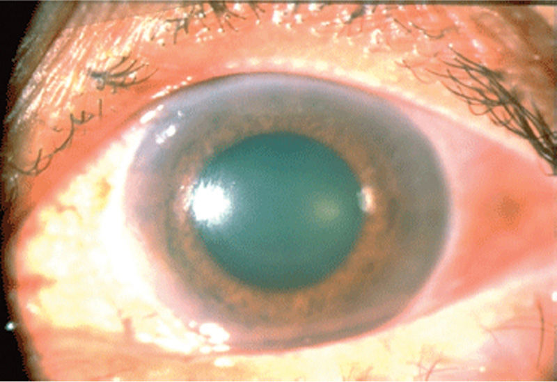
The combination of painful red eye with decreased vision, corneal edema, and a shallow anterior chamber are the findings of note seen in angle-closure glaucoma. Classic presentation of corneal abrasion is a patient who presents with the sensation of sand in the eye, and infectious keratitis is seen in patients who have recently received steroids with herpetic infections of the optics. Traumatic hyphema does have a red eye, but it occurs when a patient has frank blood in the anterior chamber (Figure 14-2).

Figure 14-2. Mid-dilated pupil and corneal clouding (seen as loss of typical corneal sheen and dulling of corneal light reflex) in a case of acute angle-closure glaucoma. (Reproduced, with permission, from McKean SC, et al. Principles and Practice of Hospital Medicine. New York: McGraw-Hill; 2012.)
Clues to diagnosis of angle-closure glaucoma:
• Red eye with pain
• Halo vision
• No history of glaucoma
Acute angle-closure glaucoma occurs most commonly at night because low light levels cause mydriasis, and folds of the peripheral iris block the angle of Schlemm.
What is the best next step in the management of this patient?
a. Pharmacologic therapy by an ophthalmologist
b. Laser iridotomy by an ophthalmologist
c. Gonioscopy by an ophthalmologist
d. Slit lamp grading by an ophthalmologist
e. Computed tomography (CT) scan of the orbit
Answer c. Gonioscopy by an ophthalmologist
The best next step in the management of this patient is to refer to an opthalmologist for gonioscopy, which is the most accurate test for diagnosing angle-closure glaucoma. This is typically combined with tonometry to garner a pressure. This uses a special lens, which allows to visualize the angle and look for closure. This must be done by ophthalmologists and is one of the few times in the entire USMLE in which a consult must be called for both diagnosis and treatment.
What is the best next step in the management of this patient?
a. Pharmacologic therapy by ophthalmologist
b. Laser iridotomy by ophthalmologist
c. Surgical iridotomy
Answer a. Pharmacologic therapy by ophthalmologist
The most appropriate therapy for angle-closure glaucoma is topical pressure-lowering eye drops combined with systemic acetazolamide. The combinations most often used for eye drops are timolol, apraclonidine, and pilocarpine. The intraocular pressure should be rechecked after 60 minutes, and if medical treatment is successful, then laser peripheral iridotomy is the next most appropriate therapy. If the angle has not opened after 60 minutes through both treatment modalities, then surgical peripheral iridectomy is the ultimate therapy.
Always check the other eye; 50% of the time, the other eye has an episode of angle closure within 5 years.
What is the most likely diagnosis?
a. Central retinal artery occlusion
b. Central retinal vein occlusion (CRVO)
c. Retinal detachment
d. Temporal arteritis
Answer b. Central retinal vein occlusion (CRVO)
CRVO occurs presumably secondary to Virchow’s triad and leads to blood flow egress compromise. Overall, it is unknown precisely why the retinal vein is obstructed. The most common location is in the central retinal vein, which causes pressure to build in the capillaries and subsequent hemorrhage and leakage of fluid and blood. Macular ischemia causes nonperfusion of the retina and, in turn, visual loss. Retinal detachment would have to be preceded by trauma, and temporal arteritis is a rheumatologic condition associated with a headache. Central retinal artery occlusion presents with a more acute painless vision loss associated with a “curtain falling over the eye” when caused by emboli.
Funduscopy will reveal dilated retinal veins.
What is the most accurate test for the diagnosis of CRVO?
a. Fluorescein angiography
b. Magnetic resonance imaging of the brain
c. Clinical examination
d. Funduscopy
Answer a. Fluorescein angiography
Fluorescein angiography of the retinal vein is most accurate test to diagnose CRVO; however, it is a test that is not always done because clinical presentation is usually enough. It is a confirmatory test when clinical examination findings are equivocal.
What is the best next step in the management of this patient?
a. Ranibizumab
b. Laser photocoagulation
c. Dexamethasone
d. All of the above
Answer d. All of the above
The treatment involves the acute administration of dexamethasone for the macular edema followed by laser photocoagulation and ranibizumab to prevent neovascularization.


