Fig. 9.1
Normal anatomy (Courtesy of Joao Paulo Kawaoka Matushita Junior, AC Camargo-Cancer Center)
Embryology and Anatomy of the Genitourinary Tract
In the 5th week of gestation, the ureteric bud (metanephric duct) becomes a branch of the Wolffian duct, next to its entry in the cloaca, which joins the metanephric blastema and becomes the primitive kidney at about the 6th week of gestation. Between the 6th and 7th weeks, the embryonic kidneys ascend, while the ureter acquires a separate opening to the bladder and migrates cranially [1].
In the 9th week of pregnancy, the kidneys are already as present as they are in adulthood. This relative upswing is basically due to the growth of the embryo’s body caudally toward the kidneys, which take their retroperitoneal position (Fig. 9.2 Normal development of kidney).

Fig. 9.2
Normal development of urogenital organs. (a) Mesonephic duct. (b) Ureteric. (c) Bud ureteric. (d) Metanephricblastema (Courtesy of Joao Paulo Kawaoka Matushita Junior, AC Camargo-Cancer Center)
In the 5th week of pregnancy, the early stages of gonadal development occurs alongside the differentiation of Leydig cells, leading to the apoptotic degeneration of paramesonephric ducts (Müllerian ducts) and the maintenance of Wolffian ducts in males. The two Müllerian ducts develop and run caudally, meeting and fusing in the midline with the growth of the urogenital sinus, eventually forming the prostatic utricle [1].
At 8 weeks of gestation, the proximal portion of each mesonephric duct becomes twisted, forming the epididymis. The remainder forms both the vas deferens and ejaculatory ducts.
The Wolffian ducts influence the development of the prostatic ducts that emerge from three areas of the epithelium and adjacent mesenchyme, in the portion of the urogenital sinus to be the floor of the prostatic urethra. Each of these groups is the source of each of the three zones of the prostate.
From the 12th week of pregnancy onward, seminal vesicles emerge as lateral outgrowths of the caudal end of each mesonephric duct. The seminal vesicles are located obliquely above the prostate, between the bottom of the bladder and the rectum; they drain into the ejaculatory ducts, which, in turn, drain into the prostatic urethra [2].
The ejaculatory ducts are tubular, bilateral structures that begin at the junction of the vas deferens and seminal vesicles and pass through the prostate into the prostatic urethra in the so-called verumontanum region [1].
Imaging Methods
The understanding of normal imaging findings on suprapubic or endorectal US, CT, and MRI is essential for understanding the obstructive causes of male infertility.
Ultrasonography (US)
US is easily accessible and is usually the first imaging study used to evaluate the genitourinary system. It is an innocuous method, with no ionizing radiation, and it enables dynamic images. However, US is one of the most operator-dependent imaging techniques. SVs are seen as symmetrical structures, with regular contours, homogeneous and hypoechoic in echotexture.
The vas deferens is seen as a slightly dilated tubular structure (ampulla) which can be seen medially to SVs. In oblique images, SVs and the terminal portion of the vas deferens can be seen together to form the ejaculatory duct [3].
The prostate gland at the base level shows more homogeneous echotexture and increased echogenicity than that of the seminal vesicles. At the midline level of the prostatic base, we often observe a hypoechoic, regular, and homogeneous area. At the glandular apex level there is the so-called “peripheral zone,” which shows homogeneous echotexture [3] (Fig. 9.3).
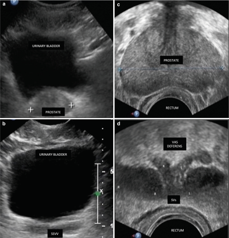
Fig. 9.3
Anatomy of the prostate, seminal vesicles, and vas deferens. (a,b) Abdominal US. (c,d) Transrectal US. Seminal vesicles (SVs) are two homogeneous, hypoechoic ovals that extend laterally upward from the bladder. Prostate is an elliptical, chestnut-shaped gland with smooth margins and a homogeneous internal echo pattern. The distal portion of the vas deferens is seen as a slightly dilated tubular structure (ampulla) medial to the SVs
Computed Tomography (CT)
A CT scan (X-ray CT) is another imaging method that can aid in diagnosis, especially of genitourinary system abnormalities. The disadvantage of CT is, nevertheless, its ionizing radiation as well as the lack of contrast in neighboring structures.
SVs are tubular, elongated, hypodense structures in a “bowtie” format, with some thin septa that can be identified after intravenous contrast [4].The prostate is presented as a hypodense image with homogeneous contrast uptake (Fig. 9.4).

Fig. 9.4
Anatomy of the prostate, seminal vesicles and vas deferens. (a,b) Axial CT image. CT noninvasive modality, identifying calcifications, soft tissue masses or cystic lesions, but radiation involved
Magnetic Resonance Imaging (MRI)
Currently, MRI has emerged as a method with excellent accuracy for the diagnosis of both acquired and congenital lesions. MRI can assess the ejaculatory duct bilaterally, from the junction of VD and SVs to the verumontanum.
SVs are seen as structures containing fluid, elongated with thin septa and low-signal intensity on T1 weighted images and high-signal intensity on T2-weighted images [4]. The intra-abdominal VD portions are seen as bilateral symmetric tubular structures with low-signal intensity on both T1- and T2-weighted images.
The prostate on T2-weighted images shows homogeneous high signal intensities in the shape of a “crescent moon” or horn on the axial plane. The prostatic capsule defines the contour of the gland and is seen as a thin hypointense line. The central, transitional, and periurethral zones are not clearly distinguished from each other by MRI; as a result, they are jointly assessed and receive the generic name of “internal gland” [5]. The normal internal gland shows intermediate signal intensity on T2 and is often interspersed with hyperintense limited foci, related to hyperplastic nodules of benign prostatic hyperplasia (BPH) in greater or smaller volume. This, in turn, is delimited by the peripheral zone structure called “surgical prostatic capsule,” which presents as a hypointense thin layer on T2, with a well-limited aspect [2, 5, 6] (Fig. 9.5).
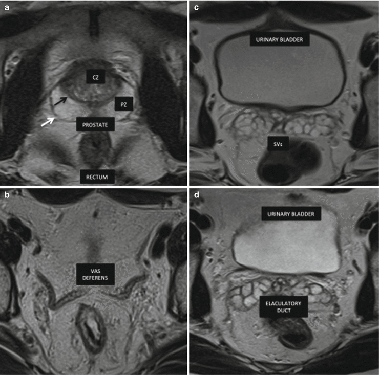
Fig. 9.5
Anatomy of the prostate, seminal vesicles and vas deferens. MRI modality of choice due to its soft tissue contrast and multi-planar capabilities and no ionizing radiation. (a) Axial T2-weighted MR image shows peripheral zone (PZ) of the prostate with homogeneous hyperitensity, and the central zone (CZ) with hypointensity of the prostate capsule (white arrow) and the surgical capsule (black arrow). (b) The vas deferens is seen as a tubular structure medial to the seminal and displays low T2 signal intensity. (c) Axial T2-weighted MR image shows the normal hyperintensity lobulated pattern of the seminal vesicles. (d) Axial T2-weighted MR image shows the anatomy of the ejaculatory duct (ED)
Development Anomalies of Vas Deferens and Seminal Vesicles
VD and SVs are urogenital subsidiary bodies, as described above. The development of these organs is related to the urinary system.
During embryogenesis, SVs or VD anomalies may be associated with renal and/or ureteral anomalies. Due to their anatomical location, SVs and VD may be involved in diseases of adjacent organs, such as the prostate and rectum [5].
SVs are responsible for most of the ejaculate; thus, agenesis or hypoplasia may lead to changes in both the volume and the quality of semen, which is, in some patients, a cause of infertility. VD tubes act as passage tubes for sperm into the ejaculatory ducts. They are also responsible for maintaining the properties of the sperm on their way to the urethra. Therefore, agenesis may, in some cases, cause changes in seminal parameters, generating infertility.
Congenital Abnormalities
Congenital Changes: Seminal Vesicles
Clinical Manifestation
In general, changes in SVs are found incidentally during imaging; in some cases, however, patients may present hematospermia, recurrent infections, and infertility.
Unilateral Agenesis of the SVs
Unilateral agenesis of the SVs occurs when there is a change in embryogenesis before the 7th week of pregnancy, when the ureteric bud originates from the mesonephric duct. It is usually associated with ipsilateral renal agenesis (79% of cases) or other renal anomalies (12% of cases), or even with normal kidney (9%) [1, 7–9] (Figs. 9.6 and 9.7).


Fig. 9.6
Agenesis of the SV and of the kidney in a 32-year-old man with infertility. CT image in the coronal (a) and axial (b) shows left renal agenesis (arrow) and ipsilateral absence of the SV (arrows)
Fig. 9.7
Agenesis of the SV and of the kidney in a 30-year-old man with infertility. CT image shows right renal agenesis (a) and absence of the SV (b) ipsilateral (arrows). Coronal (c) and axial (d) T2-weighted MR image shows again
Bilateral Agenesis of the SVs
Bilateral agenesis of the SVs is associated with gene mutations in the cystic fibrosis transmembrane conductance regulator (CFTR), causing luminal blockage by thick secretions. It is usually associated with bilateral agenesis of the VD and normal kidneys [1, 7–9] (Fig. 9.8).

Fig. 9.8
Agenesis of the SV and ED in a 26-year-old man with infertility. Axial (a) and coronal (b) T2-weighted MR image shows bilateral absence of the SV (arrows). Axial (c) T2-weighted MR image shows bilateral absence of the ED (arrows)
Hypoplasia of the SVs
Hypoplasia of the SVs refers to small congenital glands detected as imaging findings with smaller and thin septa. It may be associated with hypogonadism, cryptorchidism, and other congenital abnormalities of the genitourinary system, or be an isolated finding [1, 7–9] (Fig. 9.9)

Fig. 9.9
Bilateral SV hypoplasia in a 31-year-old man with infertility. Axial (a) and coronal, (b) T2-weighted MR image shows a bilateral small SV (arrows)
Congenital Cysts in the SVs
A congenital cyst in the SVs is usually an incidental or isolated finding. In a few cases, it is associated with ipsilateral renal agenesis, ectasia of the seminal vesicle (SV), or anomalous insertion of the ureter, known as Zinner syndrome [1, 10, 11] (Figs. 9.10 and 9.11).


Fig. 9.10
Bilateral SV cysts in a 30-year-old man with infertility. Axial (a) and coronal (c) T2-weighted MR image shows bilateral SV cysts (arrows). Axial (b) T1-weighted MR image obtained with fat saturation shows again (arrows). Axial (d) gadolinium-enhanced T1 weighted MR image shows cysts periphery enhancement (arrows)
Fig. 9.11


Zinner syndrome in 30-year-old man with infertility and hematospermia. Axial (a) T2-weighted MR image shows right renal agenesis (circle). Axial (b) T1 weighted MR image fat saturation. Note the high signal intensity within the dilated ducts, probably due to high proteinaceous/hemorrhagic content as a result of stasis (arrows). Coronal (c) T2-weighted MR image shows cystic dilatation of the seminal vesicle on the right side and right-sided ectopic ureter opening into the vas deferens inferiorly (arrows). Axial (d) T2-weighted MR image shows dilated duct that is seen indenting the posterior wall of the urinary bladder (arrow)
Stay updated, free articles. Join our Telegram channel
Full access? Get Clinical Tree
