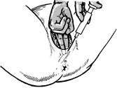
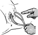
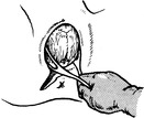
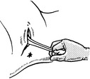
Making an incision in the perineal body at the time of delivery.
Indications
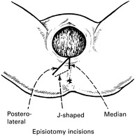
Types of Incision
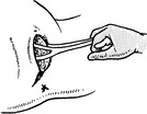
Technique

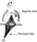
Repair
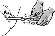
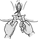
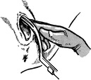
FORCEPS DELIVERY
Indications for the use of forceps
Conditions for forceps delivery
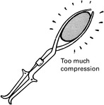
OBSTETRIC FORCEPS


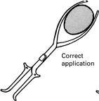
Low Forceps
Mid Forceps
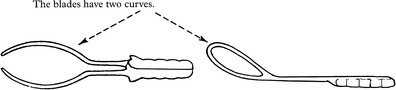
Wrigley’s Forceps

Anderson’s (Simpson’s) Forceps

Kielland’s Forceps


FORCEPS DELIVERY
Preparations
Anaesthesia
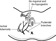
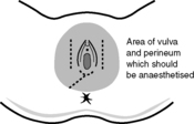
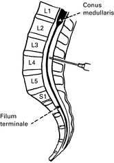
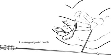
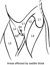
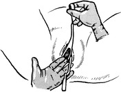
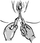
Pudendal Nerve Block
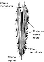
Physiology of Spinal Anaesthesia
Circulatory Effects
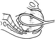
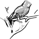
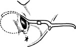
LOW FORCEPS DELIVERY


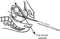
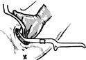
MID FORCEPS DELIVERY
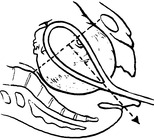
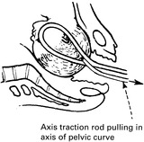
DELIVERY WITH KIELLAND’S FORCEPS


![]()
Stay updated, free articles. Join our Telegram channel

Full access? Get Clinical Tree