Chapter 10 Obstetric Hemorrhage and Puerperal Sepsis
The most common causes of maternal death are hemorrhage, embolism, hypertensive disease, and infection. In this chapter, the problems of obstetric hemorrhage and infection are considered. These conditions are associated not only with potential maternal and fetal mortality but also with significant morbidity and prolonged hospitalization.
Antepartum Hemorrhage
It is critical for the well-being of both the mother and the fetus that the patient who presents with third-trimester bleeding be evaluated and managed emergently. The differential diagnosis of third-trimester bleeding is listed in Box 10-1.
INITIAL EVALUATION
If a patient is bleeding profusely, a team approach to the assessment and management should be instituted to establish hemodynamic stability. This team should include an obstetrician, an anesthesiologist, and nurses who are knowledgeable about the management of the critically ill patient. At least two large-bore peripheral intravenous lines should be placed because this allows for the most rapid replacement of fluid and blood volume. A central venous pressure line, or preferably a Swan-Ganz catheter, is helpful in the management of hypovolemic shock.
The vital signs and amount of bleeding should be checked immediately, as should the patient’s mental status. Medical history should be checked for known bleeding disorders or liver disease, which predisposes to coagulopathy. A pelvic examination should not be performed until placenta previa has been excluded by ultrasonography. Once placenta previa has been excluded, a sterile speculum examination can be safely done to rule out genital tears or lesions (e.g., cervical cancer) that may be responsible for the bleeding. If none are identified, a digital examination may be performed to determine whether cervical dilation is present.
A complete blood count should be obtained and compared with previous evaluations to help assess the amount of blood loss, although acute blood loss may not be reflected in the hemoglobin level until homeostasis has been reestablished. An assessment of the patient’s coagulation profile should be done by obtaining a platelet count, serum fibrinogen level, prothrombin time, and partial thromboplastin time. Additionally the patient should be typed and crossmatched for at least 4 units of blood (packed cells). A rapid but subjective method to test for coagulopathy is to partially fill a red-topped tube with blood. If a clot does not form, or once formed does not stay clotted, the patient most likely has disseminated intravascular coagulopathy (DIC).
An important and accurate method for determining the cause of third-trimester bleeding is ultrasonography. This evaluation should include not only the location and extent of the placenta but also an assessment of gestational age, an estimate of fetal weight, a determination of the fetal presentation, and a screening for fetal anomalies. Uterine activity and the fetal heart rate should be assessed with a monitored strip to rule out labor and establish fetal well-being.
Abnormal Placentation
The incidence of placenta previa, the most common type of abnormal placentation, is 0.5%. Bleeding from a placenta previa accounts for about 20% of all cases of antepartum hemorrhage. Seventy percent of patients with placenta previa present with painless vaginal bleeding in the third trimester, 20% have contractions associated with bleeding, and 10% have the diagnosis made incidentally by ultrasonography or at term.
PREDISPOSING FACTORS
Factors that have been associated with a higher incidence of placenta previa include (1) multiparity, (2) increasing maternal age, (3) prior placenta previa, and (4) multiple gestation. Patients with a placenta previa have a 4% to 8% risk for having placenta previa in a subsequent pregnancy.
CLASSIFICATION
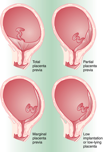
Placenta previa is classified according to the relationship of the placenta to the internal cervical os (Figure 10-1). Complete placenta previa implies that the placenta totally covers the cervical os. A complete placenta previa may be central, anterior, or posterior, depending on where the center of the placenta is located relative to the os. Partial placenta previa implies that the placenta partially covers the internal cervical os. A marginal placenta previa is one in which the edge of the placenta extends to the margin of the internal cervical os.
DIAGNOSIS
The classic presentation of placenta previa is painless vaginal bleeding in a previously normal pregnancy. The mean gestational age at onset of bleeding is 30 weeks, with one third presenting before 30 weeks. Placenta previa is almost exclusively diagnosed today by ultrasonography. Between 4% and 6% of patients have some degree of placenta previa on ultrasonic examination before 20 weeks’ gestation. With the development of the lower uterine segment, a relative upward placental migration occurs, with 90% of these resolving by the third trimester. Complete placenta previa is the least likely to resolve, with only 10% of cases resolving by the third trimester. When placenta previa is diagnosed in the second trimester, a repeat sonogram is indicated at 30 to 32 weeks for follow-up evaluation.
Transabdominal ultrasonography has an accuracy of 95% for placenta previa detection. If the placenta is implanted posteriorly and the fetal vertex is low, the lower margin of the placenta may be obscured and the diagnosis of placenta previa missed. Transvaginal ultrasonography can accurately diagnose placenta previa in virtually all cases.
MANAGEMENT
Once the diagnosis of placenta previa is established, management decisions depend on the gestational age of the fetus and the extent of the vaginal bleeding. With a preterm pregnancy, the goal is to attempt to obtain fetal maturation without compromising the mother’s health. If bleeding is excessive, delivery must be accomplished by cesarean birth regardless of gestational age. When the bleeding episode is not profuse or repetitive, the patient is managed expectantly in the hospital on bed rest. With expectant management, 70% of patients will have recurrent vaginal bleeding before completion of 36 weeks’ gestation and will require delivery. If the patient reaches 36 weeks, fetal lung maturity should be determined by amniocentesis and the patient delivered by cesarean birth if the fetal lungs are mature. Elective delivery is preferable because spontaneous labor places the mother at greater risk for hemorrhage and the fetus at risk for hypovolemia and anemia.
LOW-LYING PLACENTA
A patient with a low-lying placenta, when the placental margin is within 2 cm of the endocervical os, may present in the same way as a patient with placenta previa. It may be difficult to distinguish a low-lying placenta from a marginal placenta previa, but a transvaginal ultrasound is typically diagnostic. Although vaginal delivery is not contraindicated, the same level of monitoring should be maintained for maternal hemodynamic stability and fetal well-being.
MATERNAL-FETAL RISKS
The maternal mortality from placenta previa has dropped precipitously during the past 60 years from 30% to less than 1%. This has primarily been the result of the liberal use of cesarean delivery and careful expectant management. The rare maternal death is generally associated with complications of cesarean or uncontrolled hemorrhage from the placental site. The lower uterine segment does not contract well, especially after a lower uterine incision from cesarean delivery. DIC may also result if a massive hemorrhage or an associated abruption occurs.
The risk for antepartum or intrapartum hemorrhage, or both, is a constant threat to the patient with placenta previa. Bleeding may be exacerbated by an associated placenta accreta or uterine atony. Placenta accreta implies an abnormal attachment of the placenta through the uterine myometrium as a result of defective decidual formation (absent Nitabuch’s layer). This abnormal attachment may be superficial (accreta), or the placental villi may invade partially through the myometrium (increta) or extend to the uterine serosa (percreta). Two thirds of patients with this complication require hysterectomy. Patients with a history of uterine surgery are at greatest risk for developing an accreta. In fact, those with a prior cesarean delivery have a 25% risk.
Placenta previa predisposes to preterm delivery, which poses the greatest risk to the fetus. As a result of advances in obstetric and neonatal care, the perinatal mortality rate (PMR) for premature infants has declined over the past decade. The incidence of malpresentation with placenta previa is 30%, presumably owing to the mass effect and distortion of the lower uterine segment.
Abruptio Placentae
Abruptio placentae, or premature separation of the normally implanted placenta, complicates 0.5% to 1.5% of all pregnancies (1 in 120 births). Abruption severe enough to result in fetal death occurs in 1 per 500 deliveries.
PREDISPOSING FACTORS
Factors associated with an increased incidence of abruption are noted in Box 10-2. The most common of these risk factors is maternal hypertension, either chronic or as a result of preeclampsia. The risk for recurrent abruption is high: 10% after one abruption and 25% after two.
PATHOPHYSIOLOGY
Placental separation is initiated by hemorrhage into the decidua basalis with formation of a decidual hematoma. The resulting separation of the decidua from the basal plate predisposes to further separation and bleeding as well as to compression and destruction of placental tissue. The inciting cause of placental separation is unknown. It may be due to an inherent weakness or anomaly in the spiral arterioles. Blood may either dissect upward toward the fundus, resulting in a concealed hemorrhage, or extend downward toward the cervix, resulting in an external or revealed hemorrhage.
DIAGNOSIS AND MANAGEMENT
Clinically, the diagnosis of a placental abruption is entertained if a patient presents with painful vaginal bleeding in association with uterine tenderness, hyperactivity, and increased tone. The signs and symptoms of placental abruption are, however, variable. The most common finding is vaginal bleeding, seen in 80% of cases. Abdominal pain and uterine tenderness are present in 66% of cases, fetal distress in 60%, uterine hyperactivity and increased uterine tone in 34%, and fetal demise in 15%.
The diagnosis of placental abruption is made clinically. Ultrasonography may detect only 2% of abruptions. Because placental abruption may coexist with a placenta previa, the reason for doing an initial ultrasonic examination is to exclude the previa.
Management of the patient with an abruption includes careful maternal hemodynamic and fetal monitoring, serial evaluation of the hematocrit and coagulation profile, and delivery. Intensive monitoring of both the mother and the fetus is essential because rapid deterioration of the condition of either one can occur. Blood products for replacement should always be available, and a large-bore (16- to 18-gauge) intravenous line must be secured. Red blood cells should be given liberally if indicated. In the setting of placental abruption, the use of tocolytics or uterine relaxants is not advised. Uterine tone must be maintained to control bleeding following delivery, or at least to control the bleeding sufficiently to allow a safe hysterectomy to be performed, if necessary.
MATERNAL-FETAL RISKS
Abruption places the fetus at significant risk for hypoxia and, ultimately, death. The perinatal mortality rate due to placental abruption is 35%, and the condition accounts for 15% of third-trimester stillbirths. Fifteen percent of live-born infants have significant neurologic impairment.
Placental abruption is the most common cause of DIC in pregnancy. This results from release into the maternal circulation of thromboplastin from the disrupted placenta and subplacental decidua, causing a consumptive coagulopathy. Clinically significant DIC complicates 20% of cases and is most commonly seen when the abruption is massive or fetal death has occurred. Hypovolemic shock and acute renal failure due to massive hemorrhage may be seen with a severe abruption if hypovolemia is left uncorrected. Sheehan’s syndrome (amenorrhea as a result of maternal postpartum pituitary necrosis) may be a delayed complication resulting from coagulation within the portal system of the pituitary stalk.
Uterine Rupture
Uterine rupture implies complete separation of the uterine musculature through all of its layers, ultimately with all or a part of the fetus being extruded from the uterine cavity. The overall incidence is 0.5%.
Uterine rupture may be spontaneous, traumatic, or associated with a prior uterine scar, and it may occur during or before labor or at the time of delivery. A prior uterine scar is associated with 40% of cases. With a prior lower-segment transverse incision, the risk for rupture is less than 1%, whereas the risk with a high vertical (classical) scar is 4% to 7%. Sixty percent of uterine ruptures occur in previously unscarred uteri.
DIAGNOSIS AND MANAGEMENT
The signs and symptoms of uterine rupture are highly variable. Typically, rupture is characterized by the sudden onset of intense abdominal pain and some vaginal bleeding. Impending rupture may be heralded by hyperventilation, restlessness, agitation, and tachycardia. After the rupture has occurred, the patient may be free of pain momentarily and then complain of diffuse pain thereafter. The most consistent clinical finding is an abnormal fetal heart rate pattern. The patient may or may not have vaginal bleeding, and if it occurs, it can range from spotting to severe hemorrhage. The presenting part may be found to have retracted on pelvic examination, and fetal parts may be more easily palpated abdominally.

Stay updated, free articles. Join our Telegram channel

Full access? Get Clinical Tree

