Nutritional Dermatoses

127 Nutritional Dermatoses


Appropriate nutritional intake is an important aspect in children’s overall health. Although health care workers in developing countries are confronted daily with patients with various forms of nutritional deficiencies, these important health issues can also manifest in affluent countries with abundant food supplies. Infants and children require sufficient calories and nutrients for normal growth and development. Malnutrition in general is considered by the World Health Organization to be one of the most important risk factors for illness and premature death.


Clinical Presentation
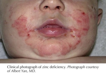


Protein-Energy Malnutrition


The majority of nutritional deficiency is associated with either poor dietary intake or GI malabsorption. Protein-energy malnutrition is divided into two major categories, marasmus and kwashiorkor. Marasmus is caused by insufficient total caloric intake and classically is seen in the context of food deprivation. Conversely, kwashiorkor is associated with inadequate protein intake in the setting of normal caloric intake. Indeed, kwashiorkor was initially described in children whose caloric intake was almost entirely derived from corn. Protein-losing enteropathies or diets consisting entirely of rice, milk because of concerns for milk intolerance are known causes of kwashiorkor in developed countries.


A child with marasmus is defined as having less than 60% of expected body weight for age in the absence of edema or hypoproteinemia. This form of malnutrition presents with loss of subcutaneous fat and muscle wasting, leading to an overall emaciated appearance. The skin is typically dry, thin, and wrinkled. Hyperpigmentation, fine scale, increased lanugo hair, nail fissures, and purpura are other cutaneous findings associated with marasmus.


Children with kwashiorkor also exhibit a lower body weight than expected for age, ranging from 60% to 80% of expected body weight. However, unlike in marasmus, these children also exhibit edema and hypoproteinemia stemming from insufficient dietary protein or the underlying medical condition leading to intestinal protein loss. Overall, the presence of mild hypoproteinemic edema in early disease gives the appearance of a well-fed, overweight child. However, recognition of the cutaneous findings help establish the diagnosis. Pigment alterations are the most common skin finding in patients with kwashiorkor, presenting as hypo- or hyperpigmentation after minor injury. In mild cases, superficial desquamation occurs that has the clinical appearance of enamel paint, which can progress in more severe cases to large areas of erosions, particularly on the extremities and buttocks, with an appearance similar to flaking paint. These areas are commonly secondarily infected with bacteria or colonized with Candida spp. Children also can present with skin atrophy, redness, and purpura. The hair is often dry, sparse, and lighter in color. Under conditions of repeated episodes of protein malnutrition followed by periods of adequate protein intake, the hair may have alternating light and dark bands, termed the flag sign, which correspond to the different episodes of protein nutritional status. Other important findings helpful in establishing the diagnosis of kwashiorkor include edema, irritability, anorexia, apathy, hepatomegaly from fatty infiltration of the liver, and failure to thrive.


Zinc Deficiency


Zinc is an element that is a required component of many enzymes involved in the synthesis and degradation of lipids, protein, and nucleic acid. Classic skin findings of zinc deficiency include erythematous and slightly eroded plaques involving the extremities, diaper area, and periorificial area. The facial involvement often involves the lower cheeks and chin but spares the skin above the upper lip, giving a “U” appearance. The rash can present with exudate, crust, vesicles, and bullae (Figure 127-1). Chronic zinc deficiency often manifests with lichenified plaques. Candida spp. and Staphylococcus aureus superinfections are common in this disorder. Other cutaneous findings of zinc deficiency include stomatitis, angular chelitis, blepharitis, nail-fold inflammation with possible nail dystrophy, and hair thinning with areas of complete alopecia. In addition to dermatitis, diarrhea is a commonly associated symptom. The severity is highly variable and does not correlate with the development of cutaneous findings. Children with zinc deficiency also characteristically are irritable, have problems eating and sleeping, and are growth impaired.


Acquired zinc deficiency is associated with poor dietary intake or underlying GI disease, leading to malabsorption. In addition to acquired zinc deficiency, an inherited cause of zinc deficiency has been identified. This disorder, termed acrodermatitis enteropathica, is caused by an autosomal recessive mutation in the zinc transporter gene SLC39A4. Because human breast milk (but not formula or cow’s milk) contains zinc-binding proteins that aid in zinc absorption, these patients typically present 1 to 2 weeks after weaning from breast milk. Interestingly, various case reports of exclusively breastfed infants with zinc deficiency demonstrated decreased zinc levels in breast milk despite normal maternal serum zinc. These infants do not exhibit a defect with intestinal zinc absorption, classifying this disorder, termed transient neonatal zinc deficiency, as an acquired zinc deficiency unlike acrodermatitis enteropathica. Transient neonatal zinc deficiency has been linked with maternal mutations in the zinc transporter gene SLC30A2, which is responsible for transport of zinc into breast milk. Understanding the genetic cause of zinc deficiency is important because the levels of zinc supplementation for acquired zinc deficiency and acrodermatitis enteropathica differ, and zinc oversupplementation can potentially result in immune dysfunction. The diagnosis of other specific nutritional deficiencies, including essential fatty acid (EFA) deficiency and biotin deficiency, should be considered when suspecting zinc deficiency because of the similarity in cutaneous manifestations of these three disorders (see below).

< div class='tao-gold-member'>

Stay updated, free articles. Join our Telegram channel

Jun 19, 2016 | Posted by in PEDIATRICS | Comments Off on Nutritional Dermatoses

Full access? Get Clinical Tree

Get Clinical Tree app for offline access