126 Nevi
Etiology And Pathogenesis
Common moles, or melanocytic nevi, are benign proliferations of melanocytes called nevus cells. They can appear tan, light brown, brown, or black in color. Nevi can be congenital or acquired. They can be typical or atypical (dysplastic). There are also other varieties of nevi such as blue nevi, halo nevi, and Spitz nevi that have some special clinical and histologic features that distinguish them from common moles.
Melanocytic nevi are extremely common in children, with almost 100% of white children developing at least one nevus by early childhood. Although most nevi are benign, trying to differentiate nevi from melanoma or nevi that have developed melanoma within them can be challenging. Melanoma is uncommon in children, with an incidence of one per million children per year. However, there has been an increase in their incidence. The Surveillance, Epidemiology and End Results (SEER) database confirms an increased incidence of approximately 2.9% per year in children from 1973 to 2001. In this database analysis, cutaneous melanoma was more common in girls (55%) and in patients older than 10 years of age (90.5%). Younger children with melanoma were more likely to be nonwhite and male. They typically presented with a primary tumor of the head and neck with regional or distant metastases. It is important to recognize and appropriately manage various types of nevi and to reduce the risk of malignant transformation.
Clinical Manifestation And Management
Common Acquired Melanocytic Nevi
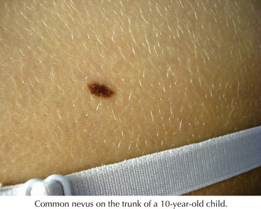
Common nevi typically begin to develop in early childhood, and new nevi may continue to arise into the third decade (Figure 126-1). These lesions have a variety of presentations depending on where the nests of nevus cells reside, and they are named accordingly. Those with nevus cells at the dermal-epidermal junction are known as junctional nevi, and those with nevus cells in the dermis are intradermal nevi. Those with cells in both compartments are called compound. The shapes and sizes vary, but most are smaller than 6 mm in size. They are more common in sun-exposed areas, where increased sun exposure leads to more moles in these areas. In general, children with lighter pigmented skin have more nevi than those with darkly pigmented skin. Children with families with increased nevi also have more moles, suggesting a genetic component to the total number of lifetime moles acquired. Studies in first-degree relatives and twins suggest that genetic factors have a significant impact on the number of moles independent of skin color or sun exposure.
The distribution of nevi can vary with skin pigmentation. Those with darker pigment have more nevi on acral locations, including the palms, soles, and nail beds. Nevi in acral sites are usually dark in color and have streaks of pigment that follow the dermatoglyphs.
Dysplastic or Atypical Nevi
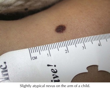
These are atypical nevi that have some asymmetry, size larger than 6 mm, variable color, and irregular borders (Figure 126-2). They frequently contain both a macular and papular component and variegated color (pink, brown, tan). They show architectural disorder histologically and are categorized by many pathologists as mildly, moderately, or severely atypical. Atypical moles typically begin to develop around puberty. The tendency to develop these nevi is genetic, with some families having multiple atypical nevi but no family history of melanoma but other families having both an increased risk of atypical moles and melanoma (familial atypical mole and melanoma syndrome [FAMM]).
Nevi that change in size, shape, color, or surface (bleeding, crusting, ulceration, thickening) or show signs of inflammation deserve evaluation and consideration for biopsy. Additionally, pain, itch, or tenderness also suggest that the nevus should be evaluated. With this said, however, symmetrical enlargement can be a normal finding in younger patients because the nevus grows with the patient. Uniform darkening after sun exposure can also occur. Chronic rubbing can also make nevi change color. Acquired nevi should be excised if they show signs of malignant transformation. They can also be removed surgically if they are in a location that is not practical for observation or if they are persistently irritated because of location such as at the waistline or neckline. All other nevi can be serially followed for change by skin examination, photography, or both.
When children have multiple moles, it is helpful to look at the clinical appearance of all of the nevi. Nevi are often very similar. This has been called the “signature” of the nevi and again highlights that nevi are programmed by genetics. A nevus that looks different from this signature should be looked at with suspicion. A new nevus is usually not concerning in a child or adolescent if it fits the signature. It is critical to perform a complete skin examination, including the scalp. Some evidence suggests that children who develop scalp nevi are at increased risk for acquiring a large number of nevi. Prophylactic removal of all nevi is not recommended because many melanomas develop de novo not from a preexisting nevus, and there is no evidence that removing all nevi decreases the risk of melanoma.

Stay updated, free articles. Join our Telegram channel

Full access? Get Clinical Tree

