81 Neuromuscular Disorders
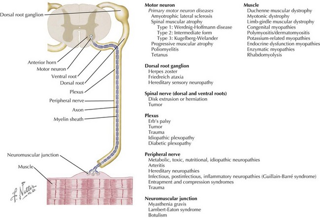
Neuromuscular disorders include a highly variable group of diseases that affect the peripheral nervous and muscular systems on any level of the neuraxis. Pathology ranges from disorders affecting the spinal motor neuron to the muscles by way of the peripheral nerves (Figure 81-1). In this chapter, several of the more common pediatric neuromuscular disorders are reviewed and classified based on their level of involvement in the neuraxis.
Anterior Horn Cell
Spinal Muscular Atrophy
The spinal muscular atrophies encompass a heterogeneous group of genetically based disorders, all of which involve a progressive degeneration of the anterior horn cells in the spinal cord and motor nuclei in the lower brain stem. The term spinal muscular atrophy (SMA) refers to the most common form, described here, but other rare forms have similar terminology (e.g., X-lined SMA). Together, they are the leading genetic cause of infant deaths, occurring in about one in 10,000 live births, with a carrier frequency of about one in 40.
Etiology and Pathogenesis
SMA is caused by an autosomal recessive mutation in the survival of motor neuron 1 (SMN1) gene on chromosome 5q13. The gene product, SMN protein, plays an important role in RNA processing and is expressed in all cells but seems to be of particular importance in motor neurons. In humans, a near-identical homocopy, the SMN2 gene, rescues an otherwise lethal disorder, and the number of copies of SMN2 is inversely related to severity of the phenotype.
Clinical Presentation
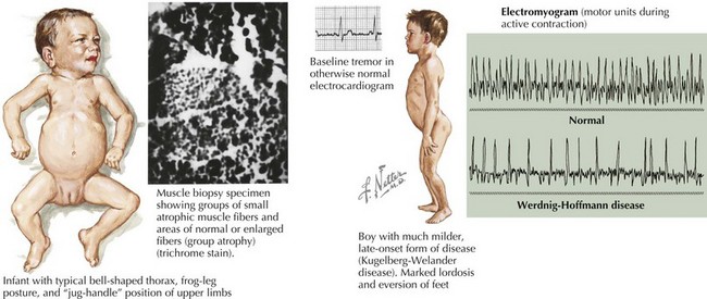
SMA is subclassified into four types based on age of onset and the maximal level of motor skills achieved. SMA type 1 (or Werdnig-Hoffmann disease) is the most common and severe of these disorders, accounting for approximately 60% of cases. SMA type 1 presents in the early infancy period (0–6 months) with generalized hypotonia, proximal and symmetric flaccid muscle weakness (initially lower more than upper limbs), and absent deep tendon reflexes. This frequently comes to the attention of the pediatrician with gross motor milestone delay and may present subacutely or more indolently. Most infants also have tongue fasciculations, and some have postural tremor of the fingers or joint contractures. Diaphragmatic sparing with intercostal muscle involvement results in paradoxical breathing and the classic bell-shaped torso (Figure 81-2). Importantly, these infants are alert and interactive with normal cognitive development and no sensory loss or impairment of eye movements. Systemic complications of SMA include pneumonia, scoliosis, poor weight gain, sleep difficulties, and joint contractures. These infants never achieve independent sitting. Most individuals’ expected life span is less than 2 years without invasive ventilatory and nutritional support.
SMA type 2 refers to infants who present usually between 6 and 18 months of age and achieve independent sitting but not ambulation and rely upon power wheelchairs for mobility. SMA type 3 presents after 18 months of age, and these children achieve community ambulation, but about half lose this ability by age 10 years. SMA type 4 presents in the adult years and tends to be slowly progressive.
The differential diagnosis includes other disorders causing acute weakness, including poliomyelitis and infantile botulism. In addition, the general differential diagnosis for hypotonia and more chronic weakness in an infant should be considered (Table 81-1).
Table 81-1 Differential Diagnosis of the Floppy Baby, Infant, and Child
Localization | Diagnoses (Examples) |
---|---|
Brain/Systemic | Chromosomal (Turner’s syndrome, trisomy 21, Prader-Willi syndrome) |
Benign congenital hypotonia | |
Infection (sepsis, meningitis, encephalitis, TORCH infections, tick paralysis) | |
Metabolic (electrolyte abnormalities, hypothyroidism, hepatic encephalopathy, mitochondrial and peroxisomal disorders, amino and organic acidemias) | |
Toxins (alcohol, narcotics, heavy metal poisoning, organophosphates, anticholinergics) | |
Neonatal encephalopathy | |
Trauma | |
Spinal cord | Hypoxic-ischemic myelopathy |
Compression | |
Syringomyelia | |
Anterior horn cell | Spinal muscular atrophy |
Infection (polio, Coxsackie) | |
Cytochrome C oxidase deficiency | |
Peripheral nerve | Demyelinating (Guillain-Barré syndrome, hereditary motor-sensory neuropathy type I, congenital hypomyelinating neuropathy) |
Axonal (familial dysautonomia, hereditary motor-sensory neuropathy type II, infantile neuronal degeneration) | |
Neuromuscular junction | Infection (botulism) |
Myasthenia gravis | |
Muscle | Myopathies and congenital muscular dystrophies |
Metabolic (acid maltase deficiency, hypo- or hyperthyroid myopathy, carnitine deficiency) | |
Muscular dystrophies | |
Inflammatory (dermatomyositis, polymyositis) | |
Mitochondrial myopathies |
TORCH, toxoplasmosis or Toxoplasma gondii, other infections, rubella, cytomegalovirus, and herpes simplex virus.
Evaluation and Management
SMA type 1 diagnosis is suspected in individuals with an appropriate clinical history and is confirmed with molecular genetic testing for homozygous deletion of the SMN1 gene. Electromyography (EMG) and nerve conduction studies (NCS) confirm a motor neuron process but is not necessary when the clinical presentation is strongly suggestive of SMA. Muscle biopsy is no longer performed as a diagnostic test. Genetic counseling and carrier testing is important after the diagnosis has been established.
There is no cure for SMA. The level of supportive care provided for SMA type 1 includes an ethical dimension, given the progressive nature of the disorder, with many parents electing to pursue a palliative course at home. Nonetheless, with aggressive management of dysphagia, malnutrition, and respiratory insufficiency, the lifespan can be extended considerably, often for several years. This entails early placement of a gastrostomy tube for supplemental feedings and early initiation of bilevel positive airway pressure (BiPAP), cough assist, and using a pump to suction oral secretions. Similar but less intensive nutritional and pulmonary support for patients with type 2 SMA, along with close attention to evolving scoliosis and joint contractures, has enabled these children to live into the third decade and beyond, often attending college, gaining employment, and forming interpersonal relationships. Children with type 3 SMA need mainly orthopedic and physical therapy support.
Future Directions
Much of the research into SMA is now focused on the molecular genetic mechanism of the disease. Several clinical drug trials based on encouraging preclinical data have been conducted on neuroprotection (riluzole), histone diacetylase (HDAC) inhibition (valproic acid), and regulators of SMN2 expression (hydroxyurea and sodium phenylbutyrate). None, however, has demonstrated clinical efficacy as yet. The use of novel oligonucleotides and small molecule drugs that increase SMN2 expression are currently in development, and stem cell and gene correction strategies are being considered.
Peripheral Nerve
Guillain-Barré Syndrome
The name Guillain-Barré syndrome (GBS) encompasses a variety of acute immune-mediated polyneuropathies. It is the most common cause of acute flaccid paralysis in infants and children in the postpolio era, with an annual incidence of 0.38 to 0.91 pediatric cases per 100,000 children. A total of 50% to 82% of pediatric cases have an antecedent respiratory or gastrointestinal (GI) infection associated with a variety of organisms, but most commonly Campylobacter jejuni (≤30% of cases). GBS has also been reported after use of several different vaccines, including influenza and rabies. There are multiple different types of GBS. Here, we will discuss acute demyelinating polyradiculoneuropathy (AIDP), which accounts for 85% to 90% of GBS cases.
Etiology and Pathogenesis
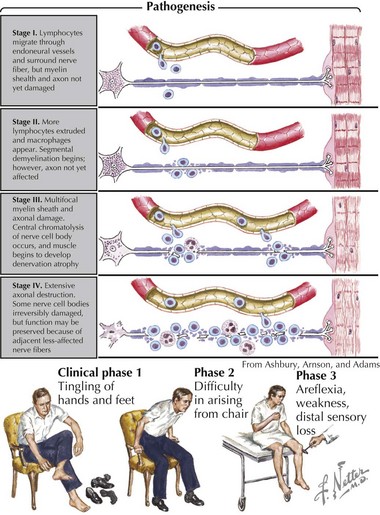
Pathologically, the two primary changes seen in GBS are acute inflammatory demyelinating polyradiculoneuropathy and acute axonal degeneration, both of which are thought to be caused by cross-reacting antibodies to the various gangliosides expressed in peripheral and cranial nerves. Campylobacter infections can result in molecular mimicry with the GM1 ganglioside, such that patients with C. jejuni infections are at 100-fold higher risk of developing GBS than the general population (Figure 81-3).
Clinical Presentation
AIDP presents 2 to 4 weeks after a respiratory or GI illness with paresthesias of the extremities and a symmetric, length-dependent weakness (beginning distally in the lower limbs) with areflexia that may ascend over hours to days. About 50% of children have autonomic dysfunction as well, including cardiac dysrhythmias, blood pressure fluctuations, or bladder dysfunction.
The differential diagnosis of GBS should include other causes of peripheral neuropathy (including toxic neuropathy, critical care neuropathy, or tic paralysis), diseases of the neuromuscular junction, or spinal cord pathology.

Stay updated, free articles. Join our Telegram channel

Full access? Get Clinical Tree

