chapter 13 Neurological Examination
As long as our brain is a mystery, the universe, the reflection of the structure of the brain will also be a mystery.
Many physicians feel uncomfortable when they are required to assess a child’s neurological status. They may be either afraid that the youngster will not cooperate or uncertain of what a “normal” child should be able to do. A competent clinical evaluation, however, often precludes unnecessary investigations and prevents unwarranted psychological and financial costs for both the patient and society.
The chapter emphasizes ways to elicit cooperation from most children so that you can determine whether the findings are significant. The nervous system is an excellent model for the logical evaluation of clinical findings. Your knowledge of neurophysiology and neuroanatomy should enable you to locate the site of the problem within the nervous system or to decide that the problem is caused by a more diffuse process and cannot be localized to a single lesion.
The neurological assessment should be stimulating and enjoyable for both you and your patient. Where relevant, the description of each part of the examination is divided according to age ranges: school-aged child, preschool-aged child, and infant. At the end of the chapter, case histories are provided to show how the assessment is applied to children of different ages and to highlight some common problems in pediatric neurology.
To avoid overlooking important findings, you should approach each phase of the neurological examination with the following categories in mind:
The Logic Behind the Approach
At each phase of the assessment, try to determine the site of the lesion. Doing so makes it easier to compile a list of differential diagnoses or hypotheses. As an initial approach, divide neurological disorders into upper motor neuron lesions (UMNLs) and lower motor neuron lesions (LMNLs). The major features of each are listed in Table 13–1. Remember that some children, such as those with leukodystrophies, may have a mixture of both UMNL and LMNL signs, and localizing the problem to a single focal lesion in such patients is not possible. In addition, children with disorders of the cerebellum or basal ganglia do not have UMNL signs.
TABLE 13–1 Signs of Upper and Lower Motor Neuron Lesions
Parameters | Upper Motor Neuron Lesions: Central Nervous System Dysfunction* | Lower Motor Neuron Lesions: Peripheral Nervous System Dysfunction† |
---|---|---|
Intellect | Deficits may be found with cortical abnormalities | Normal |
Cranial nerves | Abnormalities usually reflect brainstem involvement but may indicate a neuropathy | May be involved |
Power | Slightly decreased, although movement more severely impaired because of altered tone | Markedly reduced; neuromuscular junction disease associated with fatigue |
Tone | Increased (spasticity) with lesions affecting pyramidal pathways; rigidity seen with extrapyramidal disease | Reduced (floppy or hypotonic) |
Coordination | Impaired when cerebellum or its connections are involved | May be hindered by weakness |
Reflexes | Hyperactive in pyramidal dysfunction; plantar stimulation results in an extensor response (Babinski) in the great toe | Difficult to elicit |
Sensation | Usually intact, but spinal cord gives a sensory level that is in a dermatomal distribution | Impaired with lesions that affect the nerve |
Fasciculations | Present with anterior horn cell disease but occasionally also found with neuropathies |
* Involve intracranial contents, brainstem, or spinal cord.
† Involve intracranial horn cells, nerves, neuromuscular junction, or muscles.
Obtaining the History
When combined with a hands-off observation, the history usually provides the diagnosis (see Chapter 1). The physical examination rarely reveals previously unsuspected findings.
The school-aged child
After introducing myself to the children’s families, I try to establish a rapport with the children by asking why they think they have come visit me and what they think is wrong. Ask children their names and what they like to be called. Some children prefer to be called by a nickname.
Start the history by talking directly to the patient. This helps the child feel more involved in the process. Begin by talking about nonthreatening subjects, such as family members, school or teachers, television shows, music, or sports. Use open-ended questions to avoid “Yes” or “No” answers. All school-aged children deserve the opportunity to speak to the physician alone; it is during this part of the history that they sometimes reveal information they would otherwise withhold. The parents must be asked to leave the room for this part of the history. A child’s inability to give a history may be an important observation in itself. Having gained the child’s acceptance, you can now inquire about the problem or complaint.
Every school-aged child should be given the chance to speak to the physician alone, a time when the child sometimes reveals information that he or she would otherwise withhold. A child’s inability to give a history may in itself be an important observation.
After allowing the patient to contribute as much as he or she wishes or can, ask the parents to tell you their main concerns. First, establish whether the problem is static or progressive, a distinction that will influence your differential diagnosis and subsequent inquiries. For example, a child with learning difficulties that are worsening is much more likely to have a neurodegenerative or metabolic disorder than a child whose problems are static or nonprogressive. Similarly, an intracranial lesion, such as a tumor, is much more likely if headaches are worsening than if they have been unchanged for months. Always ask details of the child’s school performance because deterioration in grades may suggest a progressive neurological condition.
A brief but detailed history of the pregnancy is essential. Most mothers remember the pregnancy and birth in vivid detail and often have unfounded fears related to it (see Chapter 1). Reassuring a mother that the cough medicine she took during the third trimester was not the cause of her baby’s meningomyelocele may relieve anxiety. Similarly, if a child has been discharged from the neonatal unit at the same time as his/her mother, it is extremely unlikely that significant perinatal problems occurred.
The preschool-aged child
Even 2-year-old children like to be involved, and a 3-year-old child may give a more accurate history than his or her distraught parent. Start the history by talking to the patient because many children will soon become bored and distracted. The screening neurological examination of the preschool-aged child is demonstrated in two videos that are associated with this chapter (see Examination Videos 13-1 [girl] and 13-2 [boy] on Student Consult). The young child is most likely to cooperate if you frame requests in a game-like fashion. The preschool child is usually happy to show a disbelieving physician that he or she can count and identify colors. Give the child the opportunity to prove you are wrong. If you announce that Mickey Mouse is pink, even the most sullen child usually corrects you. The child’s success and your delight with his or her performance help improve the child’s confidence in a strange environment.
To have undisturbed time during the subsequent discussions with the parents, it is essential to provide the child with age-appropriate toys. This playtime also provides an excellent opportunity for you to observe the child. Rarely, it may be necessary to have the child removed to another room to play.
Observation
The school-aged child
History-taking offers an excellent opportunity to observe the child unobtrusively. The gait of a child with a hemiparesis may be apparent as he or she walks from the waiting area to the examining room. Typically, the arm is held flexed and adducted against the chest, and the leg is circumducted. The child’s appearance is very important. Note the shape and size of the head (e.g., microcephaly or hydrocephalus), the shape and positioning of the eyes and ears (e.g., in Down syndrome), and any skin lesions (e.g., the facial angioma of Sturge-Weber syndrome). Asking the parents to bring in some family photographs can sometimes be useful. This allows you to compare the child’s appearance with that of his or her relatives.
Observe the child’s gait, facial appearance, use of language, and any dysmorphic features during your first encounter.
The formal neurological examination begins during the observation phase. Strabismus or ophthalmoplegia may be apparent. The child’s use of language during interaction with the parents is often more spontaneous and informative than in conversation with the physician. Dysphasia indicates a dysfunction in the dominant hemisphere. Dysarthria may represent problems in the mouth, such as a cleft palate, or may be due to lesions that involve the cranial nerve VII, VIII, IX, X, or XII. Facial asymmetry (as in Bell palsy) may indicate a lesion of the seventh cranial nerve (LMNL) or the cerebral cortex (UMNL).
Power
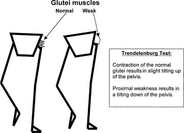
Proximal leg weakness, seen in myopathies such as muscular dystrophy, may be apparent if the child walks with a waddling gait. The Trendelenburg sign indicates a weakness of the hip abductors and is reflective of proximal weakness. Normally, when the child stands on one leg, the glutei contract, and the other side of the pelvis is tilted slightly upward. When the child has proximal weakness, the glutei are not sufficiently strong, and the other hip tilts downward. To maintain balance, the trunk leans over toward the side of the lesion; this is called the Trendelenburg sign (Fig. 13–1). Weakness and wasting of the hand muscles are more likely to indicate a neuropathy. Note that proximal weakness indicates a myopathy, and distal weakness indicates a neuropathy. Myotonic dystrophy, a dominantly inherited myopathy characterized by distal weakness and the inability to relax the muscles, is essentially the only major exception to this rule in childhood. During the period of observation, look for any asymmetry in limb movements. (See Video 13-3 on Student Consult.)

FIGURE 13–1 Trendelenburg test. Contraction of the normal gluteal muscles results in slight tilting up of the pelvis (left). Proximal weakness results in a tilting down of the pelvis (right).
Tone
A limb with increased tone (spasticity, UMNL) may be held in an unusual position. Thus, the child with a spastic arm draws with the “good” hand and keeps the spastic arm adducted against the trunk and flexed at the elbow. The spastic leg is circumducted at the hip when the child runs or walks. Always examine the wear patterns on the bottom of the child’s shoes, which give invaluable information about the child’s gait. When shaking hands with the child or parents, observe for myotonia, which is a difficulty in relaxing muscles. The most common cause of myotonia is myotonic dystrophy.
Coordination
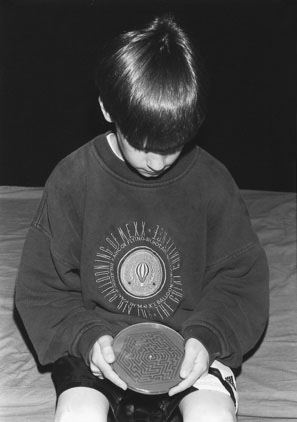
For older children, I use a challenging maze puzzle, which is an excellent test of coordination (Fig. 13–2). Cerebellar lesions affect the ipsilateral limb.
Reflexes and Sensation
Reflexes and sensation are examined during the physical examination, which is described later.
Abnormal Movements
The more common abnormal movements are described here:
The preschool-aged child
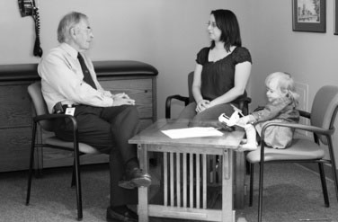
In the younger child, a period of hands-off observation should always be the first part of the physical examination. I give puzzles, a pencil, and paper to the younger child. Giving children toys allows you to observe the youngster’s manual dexterity, coordination, developmental competence, and handedness (Fig. 13–3). Observe the parent-child interaction closely. You may detect discipline problems at this stage. Drawings also reveal visual-spatial abilities, fine motor skills, and the child’s level of attention. Allowing the child to play also provides important undisturbed time with the parents as you take the history.
The infant
The period of observation of an infant is often the most revealing part of the examination. Dysmorphic features may suggest a specific syndrome. As the infant lies in his or her parent’s arms, observe the eye movements, facial movements, and symmetry. The movements of the child might indicate weakness, as is seen in anterior horn cell (AHC) disease (Werdnig-Hoffmann disease). When tone is reduced, the infant may lie with hips and arms abducted (frog-leg position). Infants may appear to be hypotonic (floppy) from either weakness (LMNL) or central causes (UMNL). With floppiness as a result of an LMNL, the infant also has significant weakness and moves very little. The infant who is floppy because of a UMNL is able to move the limbs and is not weak.
The Physical Examination
For the physical examination, the child’s cooperation is necessary and is usually easy to obtain. If a child with a behavioral problem refuses to cooperate, ask the parents to leave the room; this step is almost never necessary.
The school-aged child
I begin the physical examination of a school-aged child by playing ball with the child, thus extending the period of observation and allowing further evaluation of gait, power, and coordination as the child catches, throws, and kicks the ball.
To test proximal strength, ask the child to hop on each foot. You can also test pelvic girdle strength by asking the child to
Children with mild spasticity also have difficulty performing these tasks because of their increased muscle tone.
Test proximal strength by having the child arise from the floor without using the arms, by doing deep knee bends, and demonstrating the “duck walk.”
Next, have the child walk on the heels and then on the toes. This request is more likely to be successful if you demonstrate by walking on your heels and toes at the same time. Heel walking is an excellent test because weakness of the tibialis anterior (dorsiflexion of the foot) is an early sign of distal weakness in peripheral neuropathies. The patient with marked weakness has footdrop, which may be diagnosed from the slapping sound of the foot as the patient first walks into your office. Normally, the heel strikes the floor first, then the toes.
Milder degrees of weakness may require additional testing. To demonstrate mild proximal weakness or spasticity, you may have to ask the child to run up and down stairs. You can easily confirm mild distal problems, however, by having the child repetitively tap each foot quickly on the ground.
Next, test cerebellar function by having the child walk a straight line in a heel-to-toe fashion (tandem gait). Other useful tests of cerebellar function are the pirouette test, during which the child is expected to perform three pirouettes (360-degree turns) while walking. A variant of this test is to ask the youngster to walk several times around an object (such as a chair). In both situations, the child with cerebellar disease will stumble toward the side of the cerebellar lesion. A wide-based “drunken” gait usually indicates cerebellar disease. Tapping each foot repetitively, previously described as a test for distal weakness, is also an excellent test of coordination.
For the Romberg test, the child is told to keep the feet either side by side or in the tandem position, with one foot directly in front of the other. The child is then asked to extend the arms to the front and to close the eyes. Inability to maintain this position indicates a deficit in position sense, called proprioception. With the eyes closed, the child is deprived of visual input and must depend on proprioception to maintain the standing position. Lesions of the cerebellum result in difficulty standing, whether the eyes are open or closed. A loss of balance that occurs only when the eyes are closed is caused by a lesion in either the peripheral nerves or the posterior spinal columns.
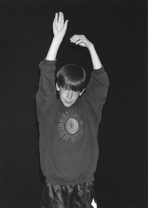
Holding the extended and supinated arms in front of the body also tests for weakness. Children with a mild weakness will be unable to maintain this position, and the arms tend to drift downward, flex at the elbow, and pronate (Fig. 13–4).
Check the spine next, while the child touches his or her toes. Look for bony deformities and midline skin lesions, such as tufts of hair or sacral dimples, which may suggest underlying malformations.
Note that up to this point, you have not laid a hand on the child or used a single strange or intimidating instrument. The entire examination has been a hands-off exercise. It is now time to produce your reflex hammer and ophthalmoscope. You should, however, already have a provisional differential diagnosis in mind, which the examination will merely confirm or refute.
At the end of the history, you should have formulated a differential diagnosis, which the examination can then confirm or refute.
If the physical examination reveals unsuspected findings, always review your history and observations to decide whether they should have led to different conclusions.
The preschool-aged child
A period of ball playing is also useful for observing preschoolers. Even a child who is reluctant to leave his or her parent’s side will usually play ball. The period of ball playing also helps establish you as an ally. You can then proceed as described for the school-aged child. Remember, however, that 4-year-olds may not successfully complete the tandem gait and that an inability to hop on one foot is not necessarily abnormal in children younger than 7 years.
You can most easily examine children younger than 6 years while they sit on a parent’s lap rather than on the examining table.
Cortical Function
School-aged and preschool-aged children
Assess each cortical area in turn.
Frontal Lobes
The posterior frontal lobes contain the motor cortex. Deficits in a frontal lobe result in signs of upper motor neuron dysfunction on the other side of the body. Motor aphasia appears in children older than 6 or 7 years if the Broca motor speech area in the dominant hemisphere is injured. The frontal lobes also control contralateral eye movements. An irritative lesion in the frontal eye fields (e.g., a seizure) causes a deviation of the eyes away from the side of the lesion; in contrast, a destructive lesion causes a deviation of the eyes toward the side of the lesion.
Disturbance of the more anterior parts of the frontal lobe may cause personality changes, irritability, and lethargy, with a lack of spontaneity. Sphincter incontinence may develop, and primitive reflexes, such as rooting, sucking, and grasp reflexes, may re-emerge. Remember that frontal lobe lesions may be difficult to detect even if they are large.
Temporal Lobes
Temporal lobe impairment may cause personality changes similar to those seen with frontal lobe damage. Language is also represented in this lobe, and lesions of the superior and middle gyri cause Wernicke aphasia, characterized by an impaired comprehension of word elements. The ability to read, write, and understand speech may be altered. If the nondominant temporal lobe is involved, the child has a distorted perception of spatial relationships and a change in musical appreciation. Test for alterations in spatial perception by asking the child to copy geometric designs. Age-appropriate designs are shown in Figure 13–5. Bilateral involvement of the hippocampus interferes with learning. Temporal lobe injury also may produce psychotic aggressive behavior. Visual symptoms are usually represented by a homonymous superior quadrantanopia.
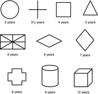
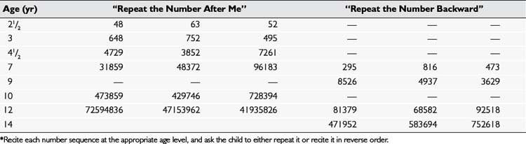
Memory deficits may be seen with a temporal lobe dysfunction. You can test a child’s immediate recall by reciting number sequences and having the child repeat them after you, either in the same order or in reverse; Table 13–2 gives examples of age-appropriate number sequences for this test.
Parietal Lobes
Parietal lobe dysfunction produces sensory perception abnormalities. Two-point discrimination, graphesthesia, and the appreciation of size, shape, and texture are all impaired. You can easily test these perceptions by asking the youngster to identify coins, a tissue, and a paper clip as you place them one at a time in one of the child’s hands while the child’s eyes are closed.
Children with parietal lobe deficits cannot appreciate simultaneous cutaneous stimulation on bilateral homologous body parts. Test this point by asking the child to identify (with the eyes closed) which arm you have touched; the child should be able to identify the simultaneous touch of both arms.
A parietal lobe injury impairs awareness on the opposite side; cortical sensory changes are best tested during the examination of sensation. Assuming that you are neurologically normal, use your own sensory perception as the normal reference.
Lesions of the parietal cortex may also cause apraxia, the inability to perform a series of tasks; apraxia may be present even though the patient can complete individually each component of the action. In young children with damage to the parietal lobes, growth on the affected side is usually impaired. Similarly, a smaller hand in a child with hemiparetic cerebral palsy indicates involvement of the parietal lobe. Such a child has reduced sensation in that limb and, therefore, has more difficulty with hand function than the child with an exclusively motor dysfunction.
Occipital Lobes
Bilateral involvement causes cortical blindness. Unilateral lesions produce homonymous visual field defects.
Cranial Nerve I (Olfactory)
The school-aged child
The olfactory nerve is responsible for the sense of smell. Your delight at a diagnosis of anosmia will tend to fade, however, if the child’s sense of smell returns after he or she blows his or her nose. Because children seem to have constant respiratory infections, first examine the nostrils. Then, while the child’s eyes are either closed voluntarily or covered by a parent’s hand, ask the child to identify the scent of common objects, such as chewing gum and chocolate, a task he or she should be able to perform. Never use irritants such as ammonia, a detection of which involves the fifth cranial nerve function.
Cranial Nerve II (Optic)
Always separately test each eye (see Chapter 8), and divide the examination into the following four parts.
Visual fields
The School-Aged Child
Ask the school-aged child to point to your wiggling finger or to report how many fingers you are extending in each of the four visual quadrants while the child looks straight into your eyes.
The Preschool-Aged Child
For the younger child, test the visual fields by distracting him or her with a toy in front while bringing a red toy from behind the youngster into the peripheral field of vision (Fig. 13–6). The child with normal vision should respond as soon as the toy reaches a line perpendicular to the outer canthus of the eye.

Stay updated, free articles. Join our Telegram channel

Full access? Get Clinical Tree

