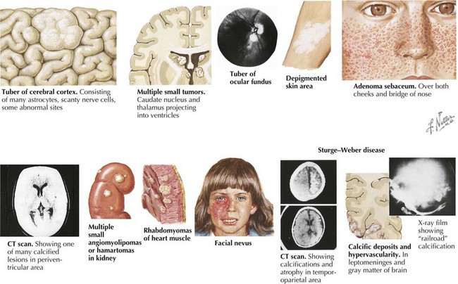
TSC is a multisystemic disorder involving the eyes, heart, lung, kidney, brain, and skin (Figure 76-1). The prevalence of TSC is one in 6000 to 10,000 live births. TSC is caused by an autosomal dominant mutation in TSC 1 on chromosome 9q34 or TSC 2 gene on chromosome 16p13. TSC can result from inherited or de novo mutations. TSC 1 and 2 code for hamartin and tuberin, respectively, which form a protein complex involved in the regulation of cell growth. In TSC, there is loss of inhibition of Rheb (a rapamycin) by TSC 1–TSC 2 complex leading to uncontrolled cell growth of hamartomas in multiple organ systems. The diagnosis of TSC is based on clinical findings, and genetic testing is corroborative. The TSC Consensus Conference in 1998 revised the diagnostic criteria (Table 76-1); a patient must have two major features, or one major and two minor features. A patient with one major and one minor feature is diagnosed with probable TSC.
Table 76-1 Criteria for Tuberous Sclerosis Complex
Major Criteria |
Minor Criteria |
|---|
Facial angiofibromas or forehead plaque |
Multiple randomly distributed dental enamel |
Nontraumatic ungual or periungual fibroma |
Hamartomatous rectal polyps |
Hypomelanotic macules (>3) |
Bone cysts |
Shagreen patch (connective tissue nevus) |
Cerebral white-matter “migration tracts” |
Cortical tuber |
Gingival fibromas |
Subependymal nodule |
Retinal achromic patch |
Subependymal giant cell astrocytoma |
Nonrenal hamartoma |
Multiple retinal nodular hamartomas |
“Confetti” skin lesions |
Cardiac rhabdomyoma (single or multiple) |
Multiple renal cysts |
Lymphangiomyomatosis |
|
Renal angiomyolipoma |
|
From Roach ES, DiMario FJ, Kandt RS, et al. Tuberous Sclerosis Consensus Conference: recommendations for diagnostic evaluation. J Child Neurol 14:401-407, 1999.
The skin lesions of TSC include hypopigmented macules (“ash leaf spots”), facial angiofibromas (adenoma sebaceum), shagreen patches, and ungual fibromas. Ash leaf spots, which are best seen with a Wood’s lamp, occur in 90% of patients and appear during infancy. Facial angiofibromas first present in preschoolers on the nose and checks and become more prominent in adolescents extending down the nasolabial folds. These skin lesions may be treated with laser therapy. The shagreen patch is a fleshy, raised lesion often located on the lower back in teenagers. Ungual fibromas, a nodular lesion underneath the nail at the cuticle, also appear in teenagers.
Cardiac rhabdomyomas occur in 50% to 70% of infants with TSC. Fetal ultrasound often detects these benign tumors. The tumors may cause dysrhythmias, such as Wolff–Parkinson-White syndrome. Rhabdomyomas are often not treated and may remit spontaneously. However, an arrhythmia may require medical management, or cardiac outlet obstruction may require surgery.
Renal manifestations of TSC include renal cysts, angiolipomas, and renal cell carcinoma (RCC). The renal cysts are detected in infants and children. They may be asymptomatic or cause hypertension or renal failure. Angiolipomas occur in approximately 75% of TSC patients older than 10 years of age. They have abnormal vasculature that is at risk for bleeding, especially if the angiolipoma is larger than 3 or 4 cm. RCC is rare in TSC patients but may occur at a younger age than the general population. Renal disease remains a significant cause of mortality in patients with TSC.
The neurologic manifestations of TSC include tubers, subependymal nodules, and giant cell tumors. Tubers are dysplastic cortical lesions resulting from disrupted proliferation, migration, and differentiation in early fetal life. Tubers are associated with the clinical triad of TSC—seizures, mental retardation, and behavior difficulties. Our appreciation of the significance of tuber count and its use in predicting prognosis has changed. It was once thought that increased tuber count correlated with a worse prognosis. However, the medical data supporting this correlation are quite limited. Current research suggests that the surrounding tissue of the tuber is epileptogenic and not the tuber itself. Subependymal nodules are hamartomas protruding from the ventricle walls. As they grow and calcify, they can obstruct the foramen of Monro, causing hydrocephalus. The symptoms and signs of hydrocephalus include lethargy, change in behavior, emesis, limited upgaze, headaches, and increased seizures. If a subependymal nodules grows to be larger than 1 cm, then it is considered a subependymal giant cell tumor. These are benign, but malignant transformation may occur. Subependymal giant cell tumors obstructing cerebrospinal fluid are surgically removed.
A total of 87% of TSC patients have retinal hamartomas, which are commonly asymptomatic. However, they may cause visual impairments, retinal detachment, or vitreous hemorrhage. The lungs in only adult-aged women may also be affected by TSC. Lymphangiomyomatosis cause shortness of breath, hemoptysis, or a pneumothorax.
Table 76-2 shows the tests and surveillance recommended for TSC.
Table 76-2 Tests and Surveillance Recommended for Tuberous Sclerosis Complex
Assessment |
Initial Testing |
Frequency of Testing |
|---|
Brain imaging: MRI or CT scan |
At diagnosis |
Every 1–3 years |
Renal ultrasound |
At diagnosis |
Every 1–3 years |
Ophthalmic examination |
At diagnosis |
As indicated |
Neurodevelopmental testing |
At diagnosis |
As indicated |
EKG |
At diagnosis |
As indicated |
ECHO |
At diagnosis |
As indicted |
EEG |
If seizures are present |
As indicted |
Chest CT |
In adulthood (women only) |
As indicated |
CT, computed tomography; ECG, electrocardiography; ECHO, echocardiography; EEG, electroencephalography.
The neurologic symptoms of TSC include epilepsy, behavior disturbances, and cognitive difficulties. Approximately 80% to 90% of patients have epilepsy. Infants with TSC are at risk for infantile spasms. The antiepileptic drug of choice for infantile spasms is vigabatrin. The goal of antiepileptic treatment is early seizure control to improve cognitive outcomes. Cognitive impairments range from mild learning disability to mental retardation. The neurobehavioral disturbances include autism and attention-deficit hyperactivity disorder.


