Chapter 13 Multifetal Gestation and Malpresentation
Multiple Gestation
Multiple gestation is defined as any pregnancy in which two or more embryos or fetuses occupy the uterus simultaneously. It is of utmost importance to recognize multiple gestation as a complication of pregnancy. Because the mean gestational age of delivery of twins is about 36 weeks, the perinatal mortality and morbidity in multiple gestation exceeds that of singletons disproportionately. Because of the additional physiologic stresses associated with two fetuses and placentas and a rapidly enlarging uterus, maternal morbidity is also increased.
ETIOLOGY AND CLASSIFICATION OF TWINNING
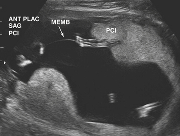
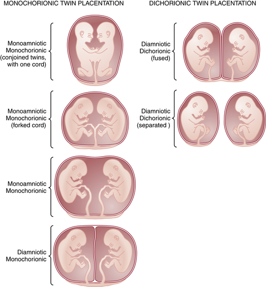
Multiple gestation occurs as the result of either the splitting of an embryo (i.e., identical or monozygotic twinning) or the fertilization of two or more eggs produced in a single menstrual cycle (i.e., fraternal or dizygotic twinning). Because dizygotic twins arise from separate eggs, they are structurally distinct pregnancies coexisting in a single uterus, each with its own amnion, chorion, and placenta. Monozygotic twins arise from cleavage of a single fertilized egg at various stages during embryogenesis, and thus the arrangement of the fetal membranes and placentas will depend on the time at which the embryo divides (Table 13-1). The earlier the embryo splits, the more separate the membranes and placentas will be. If division occurs within the first 72 hours of fertilization, the membranes will be dichorionic, diamniotic with a thick, four-layered intervening membrane. If division occurs after 4 to 8 days of development, when the chorion has already formed, monochorionic, diamniotic twins will evolve with a thin, two-layer septum. If splitting occurs after 8 days, when both amnion and chorion have already formed, the result will be monochorionic, monoamniotic twins residing in a single sac with no septum. Of all monozygotic twins, 30% are dichorionic, diamniotic, and 69% are monochorionic, diamniotic. Only 1% of twins are monoamnionic. Because twins share a sac in this type, without an intervening membrane, the risk for umbilical cord entanglement is high, resulting in a net mortality in these twins of almost 50% (Figure 13-1).
TABLE 13-1 RELATIONSHIP BETWEEN TIMING OF CLEAVAGE AND NATURE OF MEMBRANES IN TWIN GESTATIONS
Time of Cleavage∗ | Nature of Membranes |
---|---|
0-72 hr | Dichorionic, diamniotic |
4-8 days | Monochorionic, diamniotic |
9-12 days | Monochorionic, monoamniotic |
∗ Time interval between ovulation and cleavage of the egg.
INCIDENCE AND EPIDEMIOLOGY
Twins account for about 3.5% of all U.S. births. The frequency of monozygotic twinning, which depends on a very infrequent biologic event (embryo splitting), is constant in all populations studied at about 1 in 250 births. However, the frequency of dizygotic twinning, which arises from multiple ovulations in the mother, is strongly influenced by family history, ethnicity, and maternal age. A family history of dizygotic but not monozygotic twins in the maternal pedigree increases the likelihood of dizygotic twinning in subsequent generations. In western Nigeria, twinning occurs in 1 in 22 gestations, whereas in the Native American and Inuit populations, twinning is less than one fifth of that rate. Twins are twice as common in women over 35 years of age as in women at 25 years of age. Given these statistics, about two thirds of spontaneously conceived twins are fraternal, and one third are identical (monozygotic). However, in recent years, the incidence of multizygotic multifetal gestation has increased markedly with the more widespread use of ovulation induction agents and the practice of transferring multiple embryos after in vitro fertilization. The incidence of multiple gestation with the use of clomiphene is about 6% to 8%, and it is about 20% to 30% following gonadotropin therapy.
DETERMINATION OF ZYGOSITY
The prognosis and expected morbidity with twins is strongly dependent on zygosity: monozygotic twins are more likely to involve congenital anomalies, weight discordancy, twin-twin transfusion syndrome (TTTS), neurologic morbidity, premature delivery, and fetal death. Thus, determination of zygosity is the most important next step after multifetal pregnancy has been first diagnosed.
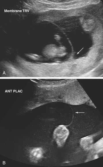
Ultrasonographic evaluation of the pregnancy is frequently very helpful in determining zygosity. Imaging of discordant fetal gender confirms a dizygotic gestation. Visualization of a thick amnion-chorion septum is suggestive of dizygotic twins, as is the presence of a “peak” or inverted “V” at the base of the membrane septum (Figure 13-2A). Conversely, in monochorionic gestation, the dividing membrane is fairly thin (Figure 13-2B). Because an early embryonic split can infrequently result in dichorionic, diamniotic twins with separate placentas, these findings are not definitive. Similarly, in rare cases of postzygotic genetic events, monochorionic twins may be gender discordant. Thus, confident diagnosis of zygosity may require detailed examination of the placenta after delivery. Thirty percent of twins will be of different sex and are, therefore, dizygotic. Twenty-three percent have monochorionic placentas and are, therefore, monozygotic. Twenty-seven percent have the same sex, dichorionic placentas, but different blood groupings, and must be, therefore, dizygotic. Twenty percent have the same sex, dichorionic placentas, and identical blood groupings. For the latter group, further studies, such as human leukocyte antigen (HLA) typing or DNA analysis, allow determination of zygosity.

FIGURE 13-2 A: Real-time ultrasound with a thick vertical amnion-chorion septum (membrane) separating one twin on the left side from the second twin on the right. The arrow points to a “peak or inverted V” suggesting dizygotic twins. B: Ultrasound of a thin vertical membrane separating one twin on the left side from the second twin on the right, suggesting a monochorionic gestational sack.
ABNORMALITIES OF THE TWINNING PROCESS
Among monozygotic multiple gestations, abnormalities in the twinning process are relatively common and include conjoined twins, interplacental vascular anastomoses, TTTS, fetal malformations, and umbilical cord abnormalities.
Conjoined Twins
If division of the embryo occurs very late (after 13 days, when the embryonic disk has completely formed), cleavage of the embryo will be incomplete, resulting in conjoined twins. Fortunately, this is a very rare event, occurring once in 70,000 deliveries. Conjoined twins are classified according to the anatomic location of the incomplete splitting: thoracopagus (anterior), pygopagus (posterior), craniopagus (cephalic), or ischiopagus (caudal). Most of such twins are thoracopagus. Delivery of conjoined twins frequently requires cesarean delivery, but postnatally, these gestations have a surprisingly optimistic prognosis in many cases. More advanced contemporary imaging has allowed detailed mapping of the shared organs and more successful surgical separation procedures.
Interplacental Vascular Anastomoses
Interplacental vascular anastomoses occur almost exclusively in monochorionic twins at a rate of 90% or more. The most common type is arterial-arterial, followed by arterial-venous and then venous-venous. Vascular communications between the two fetuses through the placenta may give rise to a number of problems, including abortion, hydramnios, TTTS, and fetal malformations. Overall, the incidence of both minor and major congenital malformations in twins is twice that in singletons, with the greater incidence of malformations occurring in monochorionic twins.
Twin-Twin Transfusion Syndrome
The presence of unbalanced anastomoses in the placenta (typically arterial-venous connections) leads to a syndrome in which one twin’s circulation perfuses the other (i.e., TTTS) in about 10% of monozygotic twins. In this syndrome, arterial blood from the “donor twin” enters the placenta (through the umbilical artery) and is taken up by the umbilical venous system belonging to the “recipient twin,” which results in a net transfer of blood from the donor to the recipient twin. Fetal complications include hypovolemia, hypotension, anemia, oligohydramnios, and growth restriction in the donor twin, and hypervolemia, hydramnios, hyperviscosity, thrombosis, hypertension, cardiomegaly, polycythemia, edema, and congestive heart failure in the recipient twin. Both twins are at risk for demise from the circulatory derangement, and the pregnancy is predisposed further for preterm delivery due to uterine overdistention with hydramnios.
TTTS is diagnosed using ultrasound. Typically the donor twin is smaller and may have oligohydramnios, absent bladder, and anemia. The recipient, on the other hand, is larger with possible polyhydramnios, cardiomegaly, and ascites or hydrops (Figure 13-3).

FIGURE 13-3 Ultrasound of a twin-twin transfusion syndrome, with one twin (upper left) in an amniotic cavity with a reduced fluid volume and a membrane (memb) separating this fetus from the second twin in an amniotic cavity with an excessive amount of fluid (right and lower half of scan image).
Given the poor prognosis of untreated TTTS (about 50% survival of either twin), treatment with either serial amniocentesis and fluid reduction from the recipient twin’s sac or laser photocoagulation of the anastomotic vessels on the surface of the placenta is performed in specialized centers.
Fetal Malformations
Arterial-arterial placental anastomoses can result in fetal structural malformations. In this situation, the arterial blood from the donor twin enters the arterial circulation of the recipient twin, and the reversed blood flow may cause thrombosis within critical organs or atresias due to trophoblastic embolization. The recipient twin, being perfused in a reverse direction with relatively poorly oxygenated blood, fails to develop normally. This so-called acardiac twin typically has no anatomic structures cephalad of the abdomen but often has fully formed lower extremities.
Umbilical Cord Abnormalities
Abnormalities of the umbilical cord occur with a higher frequency in twins and are primarily associated with monochorionic twins. Absence of one umbilical artery occurs in about 3% to 4% of twins, as opposed to 0.5% to 1% of singletons. The absence of one umbilical artery is significant because in 30% of such cases, it is associated with other congenital anomalies (e.g., renal agenesis). Marginal and velamentous umbilical cord insertions also occur more frequently in twins and may cause growth abnormalities, particularly in the third trimester.
Retained Dead Fetus Syndrome
It is not unusual for one twin to die in utero remote from term, whereas the remaining twin and the pregnancy continue to be viable. Over time (after 3 weeks or more in pregnancies that have progressed beyond 20 weeks), the retained dead fetus syndrome can develop, which involves disseminated intravascular coagulopathy in the mother as a result of transfer of nonviable fetal material with thromboplastin-like activity into her circulation. In such cases, the maternal platelet count and fibrinogen level should be checked once a week to identify possible coagulation abnormalities. The dead fetus is reabsorbed if the demise occurs before 12 weeks’ gestation. Beyond this time, the fetus shrinks and becomes dehydrated and flattened (fetus papyraceus).
ALTERED MATERNAL PHYSIOLOGIC ADAPTATION WITH MULTIPLE FETUSES
A number of normal maternal physiologic responses to pregnancy are exaggerated with multiple gestation. Whereas in normal pregnancy, maternal blood volume is augmented by 40% (2 L) over the nonpregnant baseline, in twins this increase may be 3 L or more. The increased blood volume and demand for iron and folate increase the risk for anemia in the mother and makes the patient less able to tolerate the stresses of infection, labor, and premature labor therapy. Preeclampsia and gestational hypertension are almost doubled in multifetal gestation. The increased uterine size associated with multiple fetuses can cause maternal respiratory embarrassment, orthostatic hypotension due to compression of the vena cava and aorta, and compromise of renal function due to compression of the ureters.
DIAGNOSIS
Historical factors such as a maternal family history of dizygotic twinning, the use of fertility drugs, a maternal sensation of feeling larger than with previous pregnancies, or a sensation of excessive fetal movements should raise the suspicion of twins. Physical signs, including excessive weight gain, excessive uterine fundal growth, and auscultation of fetal heart rates in separate quadrants of the uterus are suggestive but not diagnostic. An obstetric ultrasound should be performed when multiple gestation is suspected. The diagnosis of multiple gestation requires a sonographic examination demonstrating two separate fetuses and heart activities and can be made as early as 6 weeks of gestation.

Full access? Get Clinical Tree

