Chapter 6 Maternal Physiologic and Immunologic Adaptation to Pregnancy
Maternal physiologic adjustments to pregnancy are designed to support the requirements of fetal homeostasis and growth without unduly jeopardizing maternal well-being. This is accomplished by remodeling maternal systems to deliver energy and growth substrates to the fetus and to remove inappropriate heat and waste products. There appears to be a privileged immunologic sanctuary for the fetus and placenta during pregnancy.
Normal Values in Pregnancy
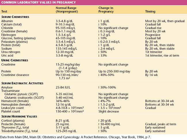
The normal values for several hematologic, biochemical, and physiologic indices during pregnancy differ markedly from those in the nonpregnant range and may also vary according to the duration of pregnancy. These alterations are shown in Table 6-1.
Cardiovascular System
CARDIAC OUTPUT
The hemodynamic changes associated with pregnancy are summarized in Table 6-2. Retention of sodium and water during pregnancy accounts for a total body water increase of 6 to 8 L, two thirds of which is located in the extravascular space. The total sodium accumulation averages 500 to 900 mEq by the time of delivery. The total blood volume increases by about 40% above nonpregnant levels, with wide individual variations. The plasma volume rises as early as the 6th week of pregnancy and reaches a plateau by about 32 to 34 weeks’ gestation, after which little further change occurs. The increase averages 50% in singleton pregnancies and approaches 70% with a twin gestation. The red blood cell mass begins to increase at the start of the second trimester and continues to rise throughout pregnancy. By the time of delivery, it is 20% to 35% above nonpregnant levels. The disproportionate increase in plasma volume compared with the red cell volume results in hemodilution with a decreased hematocrit reading, sometimes referred to as physiologic anemia of pregnancy. If iron stores are adequate, the hematocrit tends to rise from the second to the third trimester.
TABLE 6-2 CARDIOVASCULAR CHANGES IN PREGNANCY
Parameter | Amount of Change | Timing |
---|---|---|
Arterial blood pressures | ||
Systolic | ↓ 4-6 mm Hg | All bottom at 20-24 wk, then rise gradually to prepregnancy values at term |
Diastolic | ↓ 8-15 mm Hg | |
Mean | ↓ 6-10 mm Hg | |
Heart rate | ↑ 12-18 beats/min | 1st, 2nd, 3rd trimesters |
Stroke volume | ↑ 10%-30% | 1st and 2nd trimesters, then stable until term |
Cardiac output | ↑ 33%-45% | Peaks in early 2nd trimester, then stable until term |
Data from Main DM, Main EK: Obstetrics and Gynecology: A Pocket Reference. Chicago, Year Book, 1984, p 18.
Cardiac output rises by the 10th week of gestation; it reaches about 40% above nonpregnant levels by 20 to 24 weeks, after which there is little change. The rise in cardiac output, which peaks while blood volume is still rising, reflects increases mainly in stroke volume and, to a lesser extent, in heart rate. With twin and triplet pregnancies, the changes in cardiac output are greater than those seen with singleton pregnancies.
The cardiovascular responses to exercise are altered during pregnancy. For any given level of exercise, oxygen consumption is higher in pregnant than in nonpregnant women. Similarly, the cardiac output for any level of exercise is also increased during pregnancy compared with that seen in a nonpregnant state, and the maximum cardiac output is reached at lower levels of exercise. It is not clear that any of the changes in hemodynamic responses to exercise are detrimental to mother and fetus, but it suggests that maternal cardiac reserves are lowered during pregnancy and that shunting of blood away from the uterus might occur during or after exercise.
INTRAVASCULAR PRESSURES
Systolic pressure falls only slightly during pregnancy, whereas diastolic pressure decreases more markedly; this reduction begins in the first trimester, reaches its nadir in mid-pregnancy, and returns toward nonpregnant levels by term. These changes reflect the elevated cardiac output and reduced peripheral resistance that characterize pregnancy. Toward the end of pregnancy, vasoconstrictor tone, and with it blood pressure, normally increases. The normal, modest rise of arterial pressure as term approaches should be distinguished from the development of pregnancy-induced hypertension or preeclampsia. Pregnancy does not alter central venous pressures.
Blood pressure, as measured with a sphygmomanometer cuff around the brachial artery, varies with posture. In late pregnancy, arterial pressure is higher when the gravid woman is sitting compared with lying supine. When elevations in blood pressure are clinically detected during pregnancy, it is customary to repeat the measurement with the patient lying on her side. This practice usually introduces a systematic error. In the lateral position, the blood pressure cuff around the brachial artery is raised about 10 cm above the heart. This leads to a hydrostatic fall in measured pressure, yielding a reading about 7 mm Hg lower than if the cuff were at heart level, as occurs during sitting or supine measurements.
MECHANICAL CIRCULATORY EFFECTS OF THE GRAVID UTERUS
As pregnancy progresses, the enlarging uterus displaces and compresses various abdominal structures, including the iliac veins and inferior vena cava (and probably also the aorta), with marked effects. The supine position accentuates this venous compression, producing a fall in venous return and hence cardiac output. In most gravid women, a compensatory rise in peripheral resistance minimizes the fall in blood pressure. In up to 10% of gravid women, however, a significant fall occurs in blood pressure accompanied by symptoms of nausea, dizziness, and even syncope. This supine hypotensive syndrome is relieved by changing position to the side. The expected baroreflexive tachycardia, which normally occurs in response to other maneuvers that reduce cardiac output and blood pressure, does not accompany caval compression. In fact, bradycardia is often associated with the syndrome.
The venous compression by the gravid uterus elevates pressure in veins that drain the legs and pelvic organs, thereby exacerbating varicose veins in the legs and vulva and causing hemorrhoids. The rise in venous pressure is the major cause of the lower extremity edema that characterizes pregnancy. The hypoalbuminemia associated with pregnancy also shifts the balance of the other major factor in the Starling equation (colloid osmotic pressure) in favor of fluid transfer from the intravascular to the extracellular space. Because of venous compression, the rate of blood flow in the lower veins is also markedly reduced, causing a predisposition to thrombosis. The various effects of caval compression are somewhat mitigated by the development of a paravertebral collateral circulation that permits blood from the lower body to bypass the occluded inferior vena cava.
During late pregnancy, the uterus can also partially compress the aorta and its branches. This is thought to account for the observation in some patients of lower pressure in the femoral artery compared with that in the brachial artery. This aortic compression can be accentuated during uterine contractions and may be a cause of fetal distress when a patient is in the supine position. This phenomenon has been referred to as the Posiero effect. Clinically, it can be suspected when the femoral pulse is not palpable.
REGIONAL BLOOD FLOW
Blood flow to most regions of the body increases and reaches a plateau relatively early in pregnancy. Notable exceptions occur in the uterus, kidney, breasts, and skin, in each of which blood flow increases with gestational age. Two of the major increases (those to the kidney and to the skin) serve purposes of elimination: the kidney of waste material and the skin of heat. Both processes require plasma rather than whole blood, which gives point to the disproportionate increase of plasma over red blood cells in the blood expansion.
Early in pregnancy, renal blood flow increases to levels about 30% above nonpregnant levels and remains unchanged as pregnancy advances. This change accounts for the increased creatinine clearance and lower serum creatine level. Engorgement of the breasts begins early in gestation, with mammary blood flow increasing 2 to 3 times in later pregnancy. The skin blood flow increases slightly during the third trimester, reaching 12% of cardiac output.
There is little information on the distribution of blood flow to other organ systems during pregnancy. The uterine blood flow increases from about 100 mL/min in the nonpregnant state (2% of cardiac output) to about 1200 mL/min (17% of cardiac output) at term. Uterine blood flow and thus gas and nutrient transfer to the fetus are vulnerable. When maternal cardiac output falls, blood flow to the brain, kidneys, and heart is supported by a redistribution of cardiac output, which shunts blood away from the uteroplacental circulation. Similarly, changes in perfusion pressure can lead to decreases in uterine blood flow. Because the uterine vessels are maximally dilated during pregnancy, little autoregulation can occur to improve uterine blood flow.
CONTROL OF CARDIOVASCULAR CHANGES
The precise mechanisms accounting for the cardiovascular changes in pregnancy have not been fully elucidated. The rise in cardiac output and fall in peripheral resistance during pregnancy may be explained in terms of the circulatory response to an arteriovenous shunt, represented by the uteroplacental circulation. The elevations in cardiac output and uterine blood flow follow different time courses in pregnancy, however, with the former reaching its maximum in the second trimester and the latter increasing to term.
A unifying hypothesis suggests that the elevations in circulating steroid hormones, in combination with increases in production of aldosterone and vasodilators such as prostaglandins, atrial natriuretic peptide, nitric oxide, and probably others, reduce arterial tone and increase venous capacitance. These changes, along with the development of arteriovenous shunts, appear responsible for the increase in blood volume and the hyperdynamic (high-flow, low-resistance) circulation of pregnancy. The same hormonal changes cause relaxation in the cytoskeleton of the maternal heart, which allows the end-diastolic volume (and stroke volume) to increase.
OXYGEN-CARRYING CAPACITY OF BLOOD
Plasma volume expands proportionately more than red blood cell volume, leading to a fall in hematocrit. Optimal pregnancy outcomes are generally achieved with a maternal hematocrit of 33% to 35%. Hematocrit readings below about 27% or above about 39% are associated with less favorable outcomes. Despite the relatively low “optimal” hematocrit, the arteriovenous oxygen difference in pregnancy is below nonpregnant levels. This supports the concept that the hemoglobin concentration in pregnancy is more than sufficient to meet oxygen-carrying requirements.
Pregnancy requires about 1 g of elemental iron: 0.7 g for mother and 0.3 g for the placenta and fetus. A high proportion of women in the reproductive age group enter pregnancy without sufficient stores of iron to meet the increased needs of pregnancy.
Respiratory System
The major respiratory changes in pregnancy involve three factors: the mechanical effects of the enlarging uterus, the increased total body oxygen consumption, and the respiratory stimulant effects of progesterone.
RESPIRATORY MECHANICS IN PREGNANCY
The changes in lung volume and capacities associated with pregnancy are detailed in Table 6-3. Assessment of mechanical changes during pregnancy reveals that the diaphragm at rest rises to a level of 4 cm above its usual resting position. The chest enlarges in transverse diameter by about 2.1 cm. Simultaneously, the subcostal angle increases from an average of 68.5 degrees to 103.5 degrees during the latter part of gestation. The increase in uterine size cannot completely explain the changes in chest configuration because these mechanical changes occur early in gestation.
TABLE 6-3 LUNG VOLUMES AND CAPACITIES IN PREGNANCY
Test | Definition | Change in Pregnancy |
---|---|---|
Respiratory rate | Breaths/minute | No significant change |
Tidal volume | The volume of air inspired and expired at each breath | Progressive rise throughout pregnancy of 0.1-0.2 L |
Expiratory reserve volume | The maximum volume of air that can be additionally expired after a normal expiration | Lowered by about 15% (0.55 L in late pregnancy compared with 0.65 L postpartum) |
Residual volume | The volume of air remaining in the lungs after a maximum expiration | Falls considerably (0.77 L in late pregnancy compared with 0.96 L postpartum) |
Vital capacity | The maximum volume of air that can be forcibly inspired after a maximum expiration | Unchanged, except for possibly a small terminal diminution |
Inspiratory capacity | The maximum volume of air that can be inspired from resting expiratory level | Increased by about 5% |
Functional residual capacity | The volume of air in lungs at resting expiratory level | Lowered by about 18% |
Minute ventilation | The volume of air inspired or expired in 1 min | Increased by about 40% as a result of the increased tidal volume and unchanged respiratory rate |
Data from Main DM, Main EK: Obstetrics and Gynecology: A Pocket Reference. Chicago, Year Book, 1984, p 14.
As pregnancy progresses, the enlarging uterus elevates the resting position of the diaphragm. This results in less negative intrathoracic pressure and a decreased resting lung volume; that is, a decrease in functional residual capacity (FRC). The enlarging uterus produces no impairment in diaphragmatic or thoracic muscle motion. Hence, the vital capacity (VC) remains unchanged. These characteristics—reduced FRC with unimpaired VC—are analogous to those seen in a pneumoperitoneum and contrast with those seen in severe obesity or abdominal binding, where the elevation of the diaphragm is accompanied by decreased excursion of the respiratory muscles. Reductions in both the expiratory reserve volume and the residual volume contribute to the reduced FRC.
OXYGEN CONSUMPTION AND VENTILATION
Total body oxygen consumption increases about 15% to 20% in pregnancy. About half of this increase is accounted for by the uterus and its contents. The remainder is accounted for mainly by increased maternal renal and cardiac work. Smaller increments are due to greater breast tissue mass and to increased work of the respiratory muscles.
In general, a rise in oxygen consumption is accompanied by cardiorespiratory responses that facilitate oxygen delivery (i.e., by increases in cardiac output and alveolar ventilation). To the extent that elevations in cardiac output and alveolar ventilation keep pace with the rise in oxygen consumption, the arteriovenous oxygen difference and the arterial partial pressure of carbon dioxide (PCO2), respectively, remain unchanged. In pregnancy, the elevations in both cardiac output and alveolar ventilation are greater than those required to meet the increased oxygen consumption. Hence, despite the rise in total body oxygen consumption, the arteriovenous oxygen difference and arterial PCO2 both fall. The fall in PCO2 (to 27-32 mm Hg), by definition, indicates hyperventilation.
The rise in minute ventilation reflects an approximate 40% increase in tidal volume at term; the respiratory rate does not change during pregnancy. During exercise, pregnant subjects show a 38% increase in minute ventilation and a 15% increase in oxygen consumption above comparable levels for postpartum subjects.
When injected into normal nonpregnant subjects, progesterone increases ventilation. The central chemoreceptors become more sensitive to CO2 (i.e., the curve describing the ventilatory response to increasing CO2 has a steeper slope). Such increased respiratory sensitivity to CO2 is characteristic of pregnancy and probably accounts for the hyperventilation of pregnancy.
In summary, both at rest and with exercise, minute ventilation and, to a lesser extent, oxygen consumption are increased during pregnancy over the nonpregnant control values. The respiratory stimulating effect of progesterone is probably responsible for the disproportionate increase in minute ventilation over oxygen consumption.
ALVEOLAR-ARTERIAL GRADIENT AND ARTERIAL BLOOD GAS MEASUREMENTS
The hyperventilation of pregnancy results in a respiratory alkalosis. Renal compensatory bicarbonate excretion leads to a final maternal blood pH between 7.40 and 7.45. During labor (without conduction anesthesia), the hyperventilation associated with each contraction produces a further transient fall in PCO2. By the end of the first stage of labor, when cervical dilation is complete, a decrease in arterial PCO2 persists, even between contractions.
In general, when alveolar PCO2 falls during hyperventilation, alveolar partial pressure of oxygen (PO2) shows a corresponding rise, leading to a rise in arterial PO2. In the first trimester, the mean arterial PO2 may be 106 to 108 mm Hg. There is a slight downward trend in arterial PO2 as pregnancy proceeds. This reflects, at least in part, an increased alveolar-arterial gradient, possibly resulting from the decrease in FRC discussed previously, which leads to a ventilation-perfusion mismatch.
DYSPNEA OF PREGNANCY
In general, airway resistance is unchanged or even decreased in pregnancy. Despite the absence of obstructive or restrictive effects, dyspnea is a common symptom in pregnancy. Some studies have suggested that dyspnea may be experienced at some time during pregnancy by as many as 60% to 70% of women. Although the mechanism has not been established, the dyspnea of pregnancy may involve the increased sensitivity and lowered threshold to PCO2.

Full access? Get Clinical Tree

