Placenta percreta with presence of placental lacunae on ultrasound scan. Note the ‘moth-eaten’ appearance.
MRI has been shown to be equivalent to ultrasound in the diagnosis of invasive placenta [16] but may provide additional information in terms of the degree of invasion. There will be a small subset of women, especially with a posterior placenta praevia in whom invasive placenta is not apparent on ultrasound scan. MRI may provide additional information; however, neither test is fully sensitive and surgeons should proceed with caution even with negative findings on imaging.
A high index of clinical suspicion should remain in the situation of placenta praevia with previous CS. In these patients the risk of MAP is as high as 1 in 20 and in the UK the RCOG recommends preparing for the delivery of a woman with placenta praevia and one previous CS as if they had known invasive placental disease.
Antenatal Monitoring and Place of Care
Once a diagnosis has been made, a multidisciplinary discussion with the woman at the centre of care should begin. It is important for patients to be alert to bleeding and present early to hospital with even minor bleeding if outpatient management has been chosen. If managed as outpatients, patients need to be able to attend hospital rapidly and should have close vigilance at home. Prolonged inpatient admissions carry significant psychosocial morbidity for women and their families in addition to the added risk of venous thromboembolism and hospital-acquired infections. It is paramount that women and their families are fully informed of the reasons for recommending inpatient admission and participate in making the final decision in order to minimize the psychosocial harm.
Empathetic and supportive midwifery care for these women is critical as there will be many complex discussions and possibly protracted antenatal admissions that will be challenging for families preparing for birth. Women may feel cut off from the experience of ‘normal pregnancy’ and can benefit from regular one-to-one midwifery care in addition to their obstetric visits. A named midwife for each patient is the gold standard of care, so that the woman has a single contact point for questions and concerns. Ideally this midwifery team would also perform the postnatal care of these women as they would have an appreciation of the special needs of these patients.
Women who experience major haemorrhage, particularly if that leads to hysterectomy and/or critical care admission, are at risk of postnatal depression, PTSD and anxiety [17]. Therefore, they need support from an experienced team with a low threshold for referral for counselling and mental health support.
Planning the timing and place of birth should begin antenatally with the earliest suspicion of MAP, and the focus of care shifted from emergency management of massive unexpected haemorrhage to antenatal diagnosis and careful multidisciplinary planning. The multidisciplinary team should comprise experienced obstetricians, midwives, anaesthetists, neonatologists, haematologists and interventional radiologists. All units should develop a robust multidisciplinary care pathway for these patients, reflecting the interventions and expertise available in their local units.
Preoperative counselling by the surgeon should cover the choice between elective hysterectomy and uterine sparing techniques such as leaving the placenta in situ or the Triple P procedure. Some women may be interested in preserving the uterus in order to pursue future fertility. Although pregnancies have been reported after conservative management of MAP, there is a marked risk of recurrent MAP and women should be counselled regarding this, and bilateral tubal ligation (BTL) at the time of surgery should be offered. In our regional referral centre for placenta percreta, where the Triple P procedure is routinely performed, over 60% of patients have chosen to undergo simultaneous BTL. The options of cell salvage and interventional radiology either as prophylaxis or as treatment should be discussed, depending on local availability. If a patient with known invasive placenta is booked for antenatal care at a facility without these services, a transfer of care to a tertiary centre should be instituted.
If bladder invasion is known or suspected the urological surgical team should be incorporated in pre-delivery planning and may choose to participate in the delivery or site ureteric stents preoperatively. General surgery involvement is not often indicated but in the situation of known complex intra-abdominal adhesions and/or bowel involvement surgical help may be warranted.
Discussion with the anaesthetic team should include the likelihood of blood transfusion as it is estimated that over 90% of all women being delivered for MAP will need a blood transfusion and consent should be obtained for this prospectively. Women who would refuse blood should be identified and sensitively counselled by the full multidisciplinary team and a specific ‘Advance Directive’ should be prepared covering blood, cell salvage and all the available blood fractions and products. In addition, invasive monitoring via arterial and central lines should be discussed.
If delivery is planned prior to 36 weeks, the neonatology team should be involved in the pre-delivery discussions. Because of the high risk of haemorrhage if these patients labour, elective delivery at 36–37 weeks’ gestation after administration of corticosteroids may be planned. In practice, any woman with persistent vaginal bleeding or abdominal pain would be delivered at 34 weeks. Earlier elective delivery may be appropriate but should be carefully considered by the multidisciplinary team, taking into account the individual factors of each case.
The haematologist should be notified well in advance of any planned elective delivery and as soon as the decision to deliver is made in an emergency. It would be prudent to have cross-matched blood in the theatre prior to commencing surgery. Other blood products including platelets, fresh frozen plasma and cryoprecipitate should be readily available.
Management of Delivery in Patients With MAP
The delivery of patients with MAP is the subject of an NPSA/RCOG patient safety care bundle [18] comprising the minimum expected standards of care (Table 16.1). Immediately prior to delivery, regardless of the planned surgical approach, it is helpful to perform ultrasound assessment of the placental site to plan a uterine incision away from the placental site. Incision of the placenta will provoke heavy bleeding, may limit the surgical options and compromise the fetus by exsanguination.
| Consultant obstetrician planned and directly supervising delivery | |
| Consultant obstetric anaesthetist planned and directly supervising anaesthesia at delivery | |
| Blood and blood products available on-site | |
| Multidisciplinary involvement in preoperative planning | |
| Discussion and consent includes possible interventions (such as hysterectomy, leaving placenta in situ, cell salvage and interventional radiology) | |
| Local availability of level 2 critical care bed |
Peripartum Hysterectomy
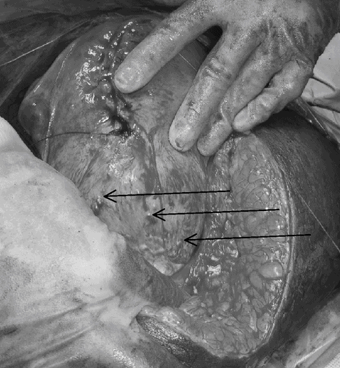
The traditional approach to MAP has been to perform a caesarean hysterectomy either as an emergency procedure in response to a massive obstetric haemorrhage or as a planned procedure. This peripartum total abdominal hysterectomy is a radical approach that may also involve resection of affected organs to remove invading placenta, for example a portion of the urinary bladder.
Elective peripartum hysterectomy has the advantage of avoiding attempts to separate the placenta, which are likely to provoke massive haemorrhage. Significantly reduced short-term morbidity (admission to intensive care unit, massive blood transfusion, coagulopathy, urological injury, return to theatre) has been demonstrated by avoiding all attempts at removing the placenta and proceeding straight to ‘elective’ hysterectomy [19]. This strategy also has the advantage of ensuring in most cases complete removal of all placental tissue at the time of delivery.
The disadvantages of peripartum hysterectomy are that bleeding may continue even after hysterectomy as the placenta may derive its blood supply from adjacent organs into which it has invaded. This may also result in damage (intentional or otherwise) to adjacent organs like the bladder, ureters or bowel into which the placenta has invaded. It also inadvertently leads to loss of fertility that may be associated with long-term psychological consequences for women.
With the advent of more conservative techniques it has become clear that hysterectomy in itself increases the psychological trauma associated with delivery, increasing blood loss, risk of ureteric and bowel injury and postoperative complications.
Uterine Conserving Measures
The main objective of conservative surgical management is to avoid the risks of intra-operative massive obstetric haemorrhage and resultant morbidity and mortality as well as avoidance of inadvertent injury to the urinary tract (or the bowel). Some women may opt for conservative surgery to preserve future fertility.
Intentional Retention of the Placenta (IRP)
Expectant management involves delivery of the fetus through an incision on the uterus above the placental border so as to avoid cutting through the placenta. In most cases, such an incision should be placed at the fundus of the uterus, in the midline and above the upper border of the placenta through a supra-umbilical midline skin incision.
Once the baby is delivered, the umbilical cord is clamped and cut very close to the placenta and ligated with an absorbable suture material. The placenta is then left inside the uterus, undisturbed. The uterine incision is closed in two layers as usual. Syntocinon is not used after the delivery of the fetus to prevent partial separation of an adherent placenta due to contraction and retraction of the uterine myometrium. Intentionally retained placenta is likely to be reabsorbed or expelled within the next 16–20 weeks. Conservative management was tried as early as 1933 because there has always been a significant demand from women for uterine sparing management options, and the evidence is building that it may in fact be physiologically less traumatic than even a planned hysterectomy.
Meyer et al. described a cohort of 12 cases of morbidly adherent placenta, which were managed by intentional retention of placenta with adjunctive interventional radiology. They used transabdominal ultrasound to identify the upper border of the placenta and the uterus was incised away from the placental site. If bleeding persisted despite inflating uterine artery balloons, the interventional radiology team performed uterine artery embolization after CS. In their series of 12 women, where interventional radiology was used with intentional retention of the placenta, only one patient needed a hysterectomy [20].
The main disadvantage of intentional retention of the placenta is the long follow-up required and the fact that as long as placental tissue is left in situ women remain at risk of secondary postpartum haemorrhage or sepsis, and may eventually undergo secondary hysterectomy for that reason. Timmermans et al. reviewed 60 case reports with successful preservation of uterus in all but 12 women, suggesting that the risk of hysterectomy in women with intentional retention of placenta is approximately 20% [21]. Antibiotics are essential and methotrexate is not recommended, as placenta at term does not have significant rapidly dividing cells.
Most clinicians recommend prolonged use of antibiotics for up to two weeks after surgery where the placenta remains in situ. Although there is little scientific evidence to support the effectiveness of this practice over the single-dose prophylaxis that is commonly used in other elective or emergency caesarean deliveries; leaving the placenta in situ theoretically increases the risk of infection. Hence, at the Regional Referral Service for Morbidly Adherent Placenta at St George’s Healthcare NHS Trust, London, antibiotics are administered for ten days for all cases of intentional retention of the placenta.
Serial ultrasound scans and βhCG may be used to document resolution of placental tissue but do not predict infection or haemorrhage. Falling levels of βhCG do not guarantee complete placental resolution and therefore measurements should be supplemented by ultrasound imaging. Although there is no scientific evidence to support the beneficial role of ultrasound for the assessment of placental involution, our experience shows that the use of colour Doppler helps to determine vascularity and may assist in assessing the overall clinical picture.
Although as previously stated, it is usual to advise women who have suffered from MAP to approach another pregnancy with caution, there are cases of future pregnancies progressing to term. In these cases, a closer antenatal surveillance as well as a planned CS should be advised.
Triple P Procedure
The Triple P procedure is a form of uterine conserving surgery bringing together a number of steps to minimize blood loss and risk of intrapartum injury to the mother [22]. It is a three-step conservative surgical alternative to peripartum hysterectomy for women with morbidly adherent placentae (Table 16.2) and early results show benefits in morbidity and maternal outcomes.
Stay updated, free articles. Join our Telegram channel

Full access? Get Clinical Tree


