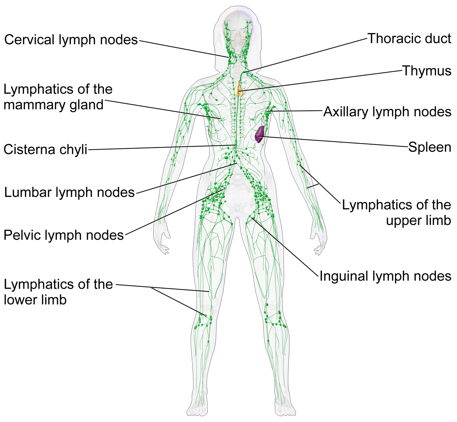
Fig. 1
The lymphatic system (Source: Bruce Blausen, Blausen Medical Communications, via Creative Commons Attributions Unported 3.0 license. https://upload.wikimedia.org/wikipedia/commons/f/f4/Blausen_0623_LymphaticSystem_Female.png. Downloaded 17 Dec 2013)
(ii)
Distribution of commonly found enlarged nodes:
1.
Head/Neck: Cervical, occipital, submental, submandibular, pre- and postauricular, clavicular.
2.
Upper extremities: Axillary, epitrochlear.
3.
Chest/Abdomen: Mediastinal, mesenteric, inguinal.
4.
Lower extremities: Femoral, popliteal.
(b)
Epidemiology: Although lymphadenopathy may present at any age, it is most common among the pediatric population. Most are benign reactive lymph nodes.
2.

Clinical features:
Get Clinical Tree app for offline access
(a)
History:
(i)
Recent illness, infections, bites, trauma, exposure to tuberculosis (TB) or new animals.
(ii)
Constitutional symptoms: fever, unintentional weight loss, night sweats, fatigue.
(b)
Physical examination: Full body examination for local or generalized enlargement of nodes.


(i)
Normal small nodes are palpable in most children.
(ii)
Examination includes palpation of spleen to detect splenomegaly and abdomen to detect enlarged abdominal lymph nodes.
(iii)
Enlarged nodes may be tender or painless, mobile or fixed, discrete, matted or, shoddy, and firm or rubbery.
(iv)
Examination of the skin surrounding the enlarged nodes may show signs of infection or trauma.
Stay updated, free articles. Join our Telegram channel
Full access? Get Clinical Tree