Genital warts.

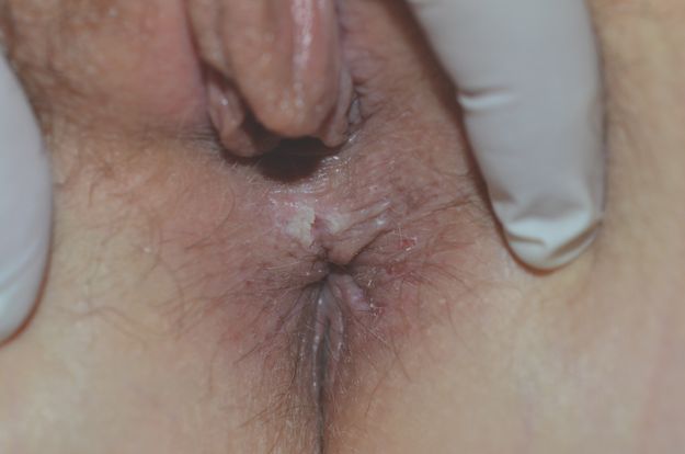
Perineal genital warts with central fissure.
The prevalence of genital warts in the adult population is about 1–2%, but many more carry HPV DNA on the external genitalia and lower genital tract. Figures range from 10 to 80% and are highly age dependent. As a result of this, people with no obvious lesions can still harbour latent virus in their anogenital epithelium and transmit it to others.
In immune-competent patients, cell-mediated immunity controls latent infection and is responsible for regression of lesions; however, in immune-compromised patients, HPV-related lesions can be persistent and are more likely to be infected with high-risk HPV types, particularly HPV-16, and to progress to neoplasia. This includes organ-transplant patients and patients with human immunodeficiency virus (HIV)/AIDS.
How Are Genital Warts Acquired?
In adults and teenagers, genital warts are considered to be a sexually transmissible infection, spread by skin-to-skin contact during intercourse. The virus is highly transmissible with up to 85% of sexual partners of patients with warts subsequently developing lesions within 6 weeks to 8 months. The incubation period is not definitely known, but the mean time is around 3 months. It is also not known how long a period of latency may exist where HPV DNA is still present on the genital area after infection, either clinical or subclinical. We simply do not know whether a patient who acquired the virus as a young person can experience a reactivation with clinical warts many years later or whether warts recurring after many years represent a new infection with another HPV type.
The use of condoms does not completely protect against the transmission of genital warts. The reason for this may be that HPV DNA is much more widely distributed on genital skin than the penis, even when there are no obvious lesions present.
It is important to remember that the patient with benign genital warts caused by non-oncogenic HPV genotypes may also harbour other potentially oncogenic types. A diagnosis of benign genital warts does not exclude the possibility of neoplasia in other parts of the lower genital tract and they should be monitored accordingly, particularly if they are immune-suppressed.
In children, however, a sexually transmissible aetiology is highly controversial, and published reports claim that there are many other ways for small children to acquire genital warts. This includes auto-innoculation, innocent transmission from other family members and vertical transmission at birth from an HPV-infected mother. There is very little logic in a medical belief system that immediately attributes genital warts in adults to sexual transmission but denies this in children.
When it comes to genital warts in children, there would seem to be a high degree of denial in the community about how they might have been acquired. The fact is, however, that many retrospective studies show that at least one in five women and one in ten men can recall sexual abuse in childhood, a sobering statistic that should make us all think critically on the subject.
Despite the current media focus on child sexual abuse within institutions, child sexual abuse within the immediate family is a well-kept secret and disclosure is a rare event. With a medical literature that throws so much doubt on the source of genital warts in small children, the best way to manage a child with genital warts becomes a significant dilemma for which there is no straightforward answer.
Presentation
Typical genital warts are raised, papillomatous, often slightly pointed lesions with a rugose surface. The warts are localised predominantly on the vulva, vaginal introitus and perianal skin but can extend into the anal canal. It is not necessary to use colposcopy to see genital warts. They are visible with the naked eye.
In general, genital warts are asymptomatic; however, when traumatised they may split or bleed, causing pain and anxiety.
Warts vary greatly in shape and size. The may be tiny and multiple, cauliflower-shaped or dome-shaped skin-coloured papules or flat-topped papules. The colour is usually skin-coloured to pink to brown and the surface dull rather than shiny. Morphology does not correlate with HPV type. Warts may be very large and numerous, particularly in the perianal area. HPV infection may also present as a fissured, painful perineal plaque that can simulate a malignancy.
The presence of genital warts is not an indication for the use of acetic acid. This stings severely on the vulva, particularly if fissuring is present, and has low specificity for the detection of HPV on skin.
HPV Vaccination
The HPV vaccine targets HPV types 6, 11, 16 and 18. It covers the two most common genotypes of benign external genital warts and the two most common genotypes associated with lower genital tract cancer. New vaccines that immunise against more genotypes will be available in the future.
Vaccination has demonstrated high-level protection against cervical dysplasia for at least 3.5 years after immunisation in adolescents aged 16–23 years, and recent data demonstrates that it has also significantly reduced the incidence of genital warts. It also promises to prevent most cases of cervical carcinoma and vulval intra-epithelial neoplasia. It must be stressed, however, that this vaccine does not stop the need for routine cancer surveillance of the cervix.
Differential Diagnosis
Because HPV is a sexually transmissible infection carrying a great degree of stigma for many patients, it is very important to be able to differentiate external genital warts from other similar lesions. If in doubt, a biopsy will usually provide the answer as warts have a typical histopathological appearance.
The main differential diagnoses are:
Seborrhoeic keratosis (see below)
Molluscum contagiosum (see below)
Vulval intra-epithelial neoplasia (see below)
Bowenoid papulosis, an unusual variant of external genital warts. In this condition, multiple domed or flat hyperpigmented papules are found on the vulva and perianal area. When biopsied, there is high-grade intra-epithelial neoplasia, similar to vulval intra-epithelial neoplasia.
Management
The appearance of genital warts does not necessarily imply infection from a new partner or infidelity in a long-term relationship. It is important to point out that an episode of genital warts may indicate a reactivation of a latent HPV infection.
There is no way to distinguish between these two situations, and so the clinician must be circumspect, and decide whether it is appropriate to advise the patient to have a full sexually transmissible infection check.
With no treatment, the natural history of genital warts is to regress spontaneously within 1–2 years. Even lesions with histopathological atypia regress. However, in some patients, HPV can be very persistent.
Genital warts are usually not symptomatic and treatment is therefore often cosmetic. Moreover, warts often recur after a single course of treatment, and there is no evidence that any treatment can change the natural history or reduce infectivity. There is no definitive first-line treatment. All treatments aim to remove visible lesions, but this does not guarantee a reduction in infectivity or eradication of HPV DNA from the anogenital skin.
A presentation with genital warts is an ideal opportunity to organise routine gynaecological screening and counselling on safe sex practices, especially for adolescents. A Pap test should always be performed at presentation to rule out significant cervical neoplasia. However, it should be pointed out that the cutaneous HPV infection that caused the warts might also cause a reversible low-grade Pap smear abnormality.
There is no evidence to indicate that treatment of visible warts reduces the risk of cancer development, or the risk of transmission. We therefore point out to patients that observation is an appropriate treatment option.
Because treatment may not change the natural history of genital warts, patients who choose treatment often require a course of therapy rather than a single treatment. In general, warts located on moist surfaces and/or in intertriginous areas respond better to topical treatment than warts on drier surfaces.
Treatments That Can Be Administered by the Patient
Many patients express a preference for applying treatment themselves. In this situation, make sure that they have been adequately counselled about how to use it and where to apply it. They should be able to contact you if adverse events (usually excessive irritation, swelling and superficial erosions) occur. In all cases, patients should take care not to apply the substances to normal skin. All treatments have a significant failure and recurrence rate, and sometimes a combination of patient-applied treatment combined with in-office treatment can be more effective than either alone.
1. Podophyllotoxin, also called podofilox and podophyllin, has been shown to be safe and effective. Patients may apply podophyllotoxin solution with a cotton swab, or podophyllotoxin gel with a finger, to visible genital warts twice a day for 3 days, followed by 4 days of no therapy. After application, it is allowed to air dry and there is no need to wash it off. This cycle may be repeated for a total of four cycles. The total wart area treated should not exceed 10 cm2, and the total volume of podophyllotoxin should not exceed 0.5 ml per application. The safety of podophyllotoxin during pregnancy has not been established.
2. Imiquimod 5% cream (Aldara® cream) is a topically active immune enhancer that stimulates production of interferon and other cytokines. Patients should apply imiquimod cream with a finger at bedtime, three times a week for as long as 16 weeks. The treatment area should be washed with mild soap and water 6–10 hours after the application. This preparation always causes an inflammatory response on treated skin, and care should be taken to avoid excessive inflammation. It therefore may not be appropriate to use imiquimod on introital skin, especially in patients predisposed to atopic skin disease. Many patients will be clear of warts by 8–10 weeks, or even sooner. The safety of imiquimod during pregnancy has not been established.
3. Green tea sinecatechins (polyphenon E ointment). This preparation is approved for use in patients over 18 years of age for external genital and perianal warts. It has been shown to be safe and reasonably effective. It is applied three times daily, leaving a thin layer on the warts. It is not necessary to wash it off.
4. Ingenol mebutate 0.015% gel and fluorouracil 5% cream. Both of these topical therapies used for actinic keratosis treatment have anecdotally been reported to be useful in eradicating genital warts. Where other treatments have failed, in a patient who is very determined to be rid of her genital warts, these treatments are a possibility. Referral to a dermatologist is advised as side effects include severe irritation.
Treatments That Are Administered by the Doctor
1. Cryotherapy with liquid nitrogen. This is suitable only for small lesions. It is important to keep the lesion frozen for 30 seconds, allow the wart to thaw and then repeat.
2. Surgical removal either by tangential scissors excision, tangential shave excision, curettage, or electrosurgery. Surgical removal of warts has an advantage over other treatment modalities in that it renders the patient wart free, usually with a single visit. This is particularly appropriate in cases where the lesions are very numerous and are interfering with function, particularly in the perianal area. In children, a general anaesthetic is usually required.
3. Surgical laser ablation with a CO2 laser. This has similar application to surgical treatment; however, there is a risk to the operator from aerosolised HPV DNA, and clinicians who do this treatment should be appropriately gowned and gloved.
4. Cidofovir. This is an antiviral drug with activity against a broad spectrum of DNA viruses including HPV. It has been shown to be effective when used topically by the patient as a 1% cream or gel, or intra-lesionally by the doctor. This use is off label and the cost is considerable. There is a growing body of evidence that this treatment is useful for anogenital warts in adults and children, and it may be useful in future.
External Genital Warts in Pregnancy
Genital warts can become much more severe in pregnant women but usually improve after the baby is born. Many experts advocate their removal at this time. HPV-6 and -11 can cause laryngeal papillomatosis among infants, and the route of transmission (transplacental, birth canal or postnatal) is not completely understood. The preventative value of caesarean delivery is unknown; thus, caesarean delivery should not be performed solely to prevent transmission of HPV infection to the newborn.
Cryotherapy is safe in pregnancy; however, topical treatments are contraindicated.
Follow-Up
After visible genital warts have cleared, patients should be cautioned to watch for recurrences, which occur most frequently during the first 3 months.
Treatment of Sex Partners
Examination of sex partners is not necessary for management of genital warts because the role of reinfection is probably minimal. Therefore, treatment to reduce transmission is not necessary. Because treatment of genital warts does not eliminate HPV colonisation, patients should be cautioned that they may still be at risk of wart recurrence.
Psychological Issues
The emotional impact of genital warts is huge. Many patients say they feel ‘dirty’, and this is usually why they seek treatment for a condition that is asymptomatic. The discovery of warts for a woman in a monogamous relationship may place great stress on the relationship. Because of the unknown incubation period, it is very difficult to be categorical about the source of the warts, and there is often never a satisfactory answer. Couples may need counselling to come to terms with the diagnosis.
Molluscum Contagiosum
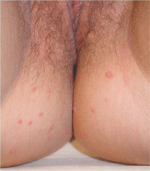
Molluscum contagiosum (Figure 7.3) is a viral infection that is very common in children but less so in adults. When it occurs on the vulva in adults, it is usually sexually acquired, but in children it is more common and is usually acquired from swimming pools and siblings with whom they share a bath. Auto-inoculation is also an important method of transmission. In children, genital mollusca are rarely found in isolation without evidence of more widespread infection of other parts of the skin.

Molluscum contagiosum. Note the typical central umbilication.
There are four genotypes of molluscum contagiosum virus (MCV), and studies show that MCV-1 is the type usually found in children, while MCV-2 is found in adults with sexually transmissible infection.
Presentation
Following an incubation period of 2 weeks to several months, the mollusca are seen as umbilicated flesh-coloured papules of 2–5 mm with a pearly appearance. Giant mollusca can occur on the genital region, and occasionally they may become inflamed and superinfected. Very small lesions often lack typical umbilication.
Mollusca of the genital area are usually asymptomatic, but itch and a secondary dermatitis may occur, particularly in atopic children. Mollusca can be severe in immunosuppressed patients, but in a healthy patient, severe mollusca is not necessarily a sign of any other disease. Some patients are simply more susceptible than others.
Mollusca are self-limiting within 6 months to 2 years in immunocompetent patients but may have a prolonged course in immunosuppressed patients. After they resolve, they are unlikely to recur.
Investigation
Diagnosis is usually made clinically; however, in the genital area, it may occasionally be difficult to differentiate small mollusca without typical umbilication from genital warts.
If there is any doubt, the diagnosis can be confirmed by microscopy performed on material from a lesion obtained by extruding the central core with pressure, a small curette or a simple shave biopsy. Histopathology is highly characteristic.
Management
Treatment of perianal and perigenital mollusca can be difficult in small children but is very easy in adults. Avoidance of baths and swimming pools may reduce auto-inoculation. If pruritus is a problem, topical corticosteroids are helpful; however, topical immunomodulators should be avoided, as both pimecrolimus and tacrolimus have been implicated in the spread of the infection. Unless the lesions are distressing, it may be best to allow them to resolve spontaneously.
Mollusca lesions have a small round core containing viral particles. If this is removed or damaged, the lesions resolve quickly.
Methods that work for removing mollusca include the following:
Scrape them off gently with a small skin curette
Squeeze the lesions to remove the core
Prick with a needle to flick out the core
Lightly freeze with a cryotherapy unit
Imiquimod (Aldara®) has been recommended as a treatment for mollusca. However, it is expensive, irritating and not completely reliable.
Cantharidin is a topical substance that causes blistering. It can be useful in mollusca in children; however, use on the genital area would be very irritating and is better avoided.
In adults the best course of action is physical removal. In children leaving them to resolve spontaneously while reducing auto-innoculation by substituting showers for baths is usually more appropriate.
Seborrhoeic Keratoses
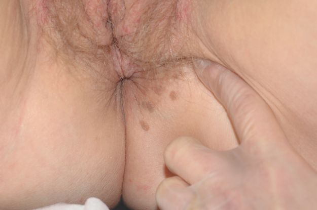
These very common benign skin lesions are common in people over the age of 40 years (Figure 7.4). They can be found on any part of the skin including the vulva. They do not occur in the vagina.

Seborrhoeic keratoses.
Presentation
Seborrhoeic keratoses have very varied shapes, sizes and colours. They may be flat, raised or pedunculated and often closely resemble warts. Colours range from skin tone to black. They may thus be confused with melanoma. They become more numerous with age.
These lesions are usually not symptomatic but if large may rub on clothing and become irritating. If they are scratched or traumatised, they may suddenly enlarge and darken.
Investigation
The main significance of seborrhoeic keratoses on the vulva is their resemblance to genital warts and sometimes to vulval intra-epithelial neoplasia (VIN; also called squamous cell carcinoma in situ). However, they are easily differentiated on biopsy as they have a typical histological appearance.
Dermoscopy is also frequently diagnostic of seborrhoeic keratoses, showing classical follicular plugging. However, it is awkward to perform on the vulva without a set-up that allows a dignified distance between the doctor and patient. This can be achieved by using a dermatoscope attached to a camera.
Management
Treatment of these lesions can be successfully achieved with:
Cryotherapy
Light curettage and cautery
Excision of large lesions
Recurrences may occur but are less common than with external genital warts.
Skin Tags (Fibromas)
These harmless lesions are common in the major flexures. They are most often found in the axillae, neck and inguinal folds.
Skin tags usually form in middle age and can occur during pregnancy. The appearance of multiple skin tags in young people is unusual and should prompt a search for signs of tuberous sclerosus, neurofibromatosis or, if perianal, Crohn’s disease.
Skin tags are small, soft, pedunculated lesions, flesh coloured to brown in colour. They often accompany seborrhoeic keratoses. The base of the peduncle is rarely wider than 1–2 mm.
Management
There is no medical need to treat skin tags, but many patients request removal because they rub on clothing.
The easiest treatment is scissor amputation at the base of the peduncle. If this is done quickly, no local anaesthetic is required unless they are quite large. Bleeding can be stopped with pressure.
Any destructive method will work: hyfrecation or cryotherapy is also effective.
Vulval Papillomatosis
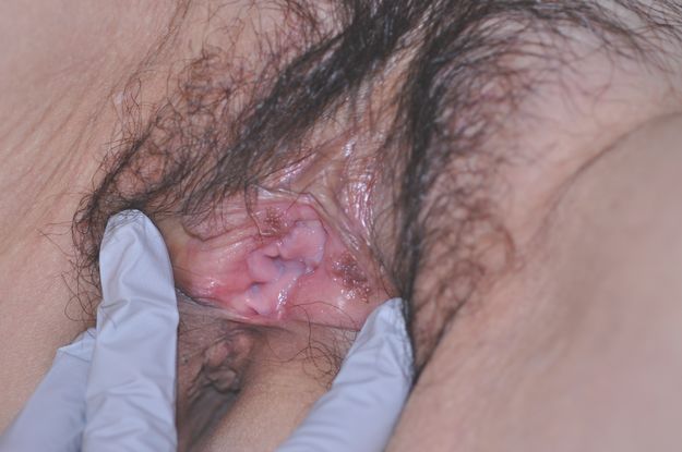
This is an unusual but important condition because it is very likely to be mistaken for genital warts. Indeed, in the past, the medical literature has suggested that vulval papillomatosis is an HPV-related condition. This is incorrect. Vulval papillomatosis is a normal variant (Figure 7.5).

Vulval papillomatosis.
Stay updated, free articles. Join our Telegram channel

Full access? Get Clinical Tree


