Author
N
Comparators
Recurrence rate (%)
Mathevet et al. [10]
86
Cold knife cone
3.6
LEEP
6.9
Laser
6.9
Mitchell et al. [11]
390
LEEP
13
Cryotherapy
19
Laser
13
Alvarez et al. [12]
375
LEEP
7
Laser
4
Singh et al. [13]
200
LEEP
6
Cryotherapy
12
Chirenje et al. [14]
400
LEEP
3.6
Cryotherapy
11.7
In a study by Saidi et al., operative variables of office-based LEEP procedures were compared with those for cold knife cones performed in the operating room. This study showed that LEEPs had significantly less blood loss (mean of 40 cm3 vs. 195 cm3), shorter operative time (mean of 5 min with a maximum of 8 min vs. a mean of 22 min), and less postoperative pain (all women undergoing cold knife cone required narcotic medications while women who underwent a LEEP required only naproxen). Furthermore, no patient undergoing a LEEP was hospitalized postoperatively, postoperative bleeding was significantly reduced, and the cost of the procedure was significantly less (mean $550 vs. $2520). These significant differences from the cold knife cone procedure make this a reasonable option for an office-based procedure, and in fact, during this study, the LEEP procedure was successfully moved from a hospital-based procedure to an office-based procedure [17].
Necessary Equipment (Table 16.2)
Table 16.2
LEEP equipment and procedure steps
Equipment | Steps of procedure |
|---|---|
Coated speculum | Obtain patient consent |
Smoke evacuation system | Position patient in dorsal lithotomy position and place return electrode |
Large cotton swabs | Place speculum and maximize separation of vaginal walls |
3–5 % acetic acid solution and/or Lugol’s solution | Anesthetize cervix |
Colposcope | Choose loop size and select power settings |
Lidocaine injection or spray | Activate smoke evacuation system |
LEEP machine with grounding pad and electrode | Perform ectocervical excision and orient specimen for pathology |
Multi-sized loop electrodes and ball electrode | Change to smaller loop and perform endocervical excision (if indicated) |
Endocervical curettes | Change to ball electrode and change settings on machine |
Smooth forceps | Cauterize LEEP bed and edges of ectocervix |
Monsel’s solution | Apply Monsel’s (as needed) |
Suture material | Give discharge instructions |
Vaginal packing (curlex) |
Coated speculum
Smoke evacuation system
Large cotton swabs
3–5 % acetic acid solution and/or Lugol’s solution
Colposcope
Lidocaine injection (see Table 16.3 for warnings) or spray
Table 16.3
Lidocaine injection warnings
Pain at injection site
Ringing in ears
Tingling at the tip of the tongue or funny taste in the mouth
Transient heart “racing”
LEEP machine with grounding pad and electrode
Multi-sized loop electrodes and ball electrode
Endocervical curettes
Smooth forceps
Monsel’s solution
Suture material
Vaginal packing (such as Kerlex)
Step by Step of the Procedure (Table 16.2)
Prior to the LEEP, the procedure must be explained including all risks and benefits, and consent should be signed. Additionally, a pregnancy test should be performed and confirmed to be negative, and the physician should ensure that the patient does not have any overt symptoms of vaginitis. If a pregnancy test returns positive, then the physician will need to reevaluate if the procedure is still indicated; if so, additional risks must be discussed with the patient. If the patient does have overt signs of vaginitis, she can be tested and treated, and LEEP can be rescheduled following treatment.
Once consent is signed and pregnancy test is confirmed negative, the procedure can be started. No prophylactic antibiotics are indicated for this procedure, regardless of valvular conditions or prosthetics. The patient is asked to move into the appropriate position, placing her feet into stirrups and lying back into the dorsal lithotomy position. Once positioned appropriately, with her buttocks hanging over the edge of the exam table, the coated speculum is inserted into the patient’s vagina.
Next, the cervix is evaluated by colposcopy. Large cotton swabs are used to apply 3–5 % acetic acid solution and/or Lugol’s solution to the cervix, and a colposcope is then used to visualize the cervix and identify any acetowhite changes or areas of nonstaining in patients evaluated with Lugol’s. The squamocolumnar junction is also identified.
A local anesthetic may then be applied to the cervix. Lidocaine with epinephrine may be used to administer a paracervical block or lidocaine spray may be utilized. This can be performed either by injecting in 2 or 4 places on the cervix. We typically utilize a total of 3–5 mL of 2 % lidocaine with 1:100,000 epinephrine injected at 2, 4, 8, and 10 o’clock.
In order to perform the electro-excision procedure, a grounding pad must be placed on the patient’s thigh and the appropriate loop electrode must be selected. The electrosurgical generator should be used in a blend mode (often 80 % cutting 20 % coagulation).
The loop is then carefully positioned close to the cervix. The LEEP may be performed in either a vertical or horizontal fashion. If, for example, the lesion is wide and narrow, it may be best to proceed from right to left (or left to right if the practitioner is left handed). If the lesion is more vertically aligned, then the practitioner can begin the LEEP at the 12 o’clock position on the cervix (at the uppermost part of the lesion) and carry the electrode down to the 6 o’clock position on the cervix.
Prior to touching the cervix, the loop electrode should be activated, and once activated, the energy should not be stopped until the loop electrode has exited the cervical tissue. A LEEP should therefore be performed in one continuous motion. Once activated, the loop should be moved directly into the cervix to the desired depth (Fig. 16.1). Once at the desired depth, the loop should be carried down or across the cervix (at the same depth) until the most distant desired area has been reached. Then the loop should be pulled directly out of the cervical tissue. The LEEP is then complete, and the specimen can be removed with a smooth forceps. An ECC should be performed after the removal of the specimen to ensure that there is no remaining lesion higher up in the endocervical canal.
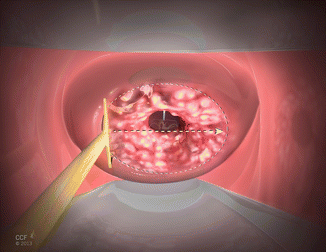
Fig. 16.1
Basic LEEP procedure. The loop electrode is moved steadily across the cervix, here right to left, to excise the abnormal region and transformation zone
The cervical bed should be carefully inspected to ensure that there is no bleeding from the site. A 3 or 5 mm ball attachment can be used to cauterize the crater and the edges of the lesion. Electrosurgical settings should be changed to cautery mode for this part of the procedure, and can be set on a range from 50 to 80 W. Pure coagulation mode should be utilized for hemostasis. Monsel’s solution may alternatively be used on the cervix or can be used in addition to ball cautery as needed to achieve hemostasis and prevent posttreatment bleeding.
Post-treatment Instructions for Patients
Patients are instructed to refrain from strenuous activity for 24–48 h and not to place anything in the vagina for 4 weeks. Tylenol or nonsteroidal anti-inflammatory medications are typically used for discomfort. Patients are instructed to call if heavy vaginal bleeding ensues or if a fever greater than 101.0 °F is noted, especially if related to increasing pelvic pain or vaginal drainage (Table 16.4).
Table 16.4
Post-LEEP instructions
Refrain from strenuous activity for 24–48 h |
Nothing in the vagina for 4 weeks |
Tylenol or nonsteroidal anti-inflammatory medications for discomfort |
Call the doctor’s office for: |
• Heavy vaginal bleeding
Stay updated, free articles. Join our Telegram channel
Full access? Get Clinical Tree
 Get Clinical Tree app for offline access

|