CHAPTER 10 LIVER PATHOLOGY


HANDLING OF THE LIVER BIOPSY SPECIMEN
• Detailed clinicopathological correlation is essential in order to obtain maximum information from the liver biopsy

• Biopsy processing and handling is generally similar to adult liver biopsies but may require additional / different testing


PROLONGED NEONATAL CHOLESTASIS
INTRODUCTION


• Many of these diagnoses can be made or suspected on the basis of diagnostic imaging, biochemical, microbiological, virological and genetic investigations

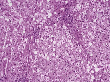
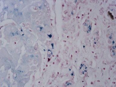
• Striking iron overload may be a non-specific feature in a liver biopsy from a neonate with liver failure
• In infants who have been treated in neonatal intensive care units, jaundice due to episodes of sepsis and prolonged total parenteral nutrition may render interpretation of the diagnostic changes in a liver biopsy difficult

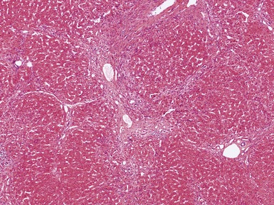
EXTRAHEPATIC BILIARY ATRESIA


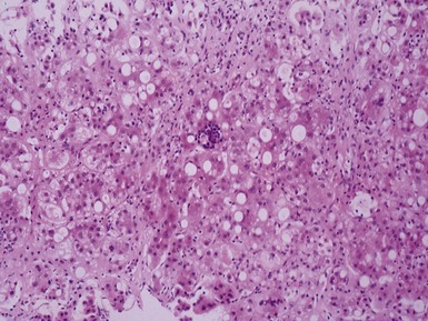
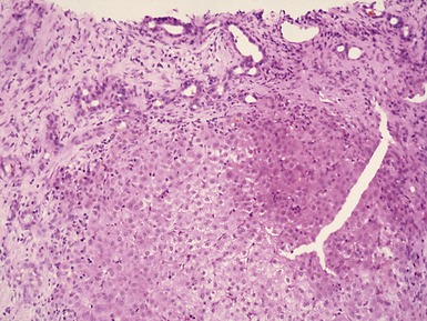
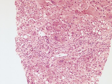
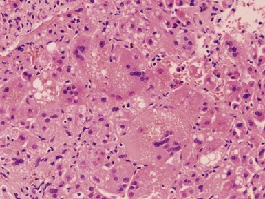
Histopathological features (Figs 10.1–10.3)


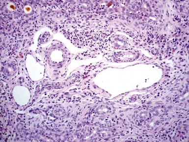
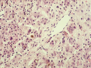
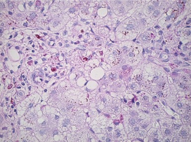
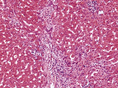
Fig 10.2 Photomicrograph of liver parenchyma demonstrating cholestasis and a small cholangiolar bile plug.
Differential diagnoses and pitfalls


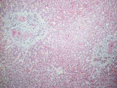
• Inspissated cholangiolar bile plugs and paucity of interlobular bile ducts are most useful features in early biopsies

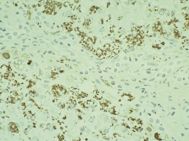
• Immunostaining for low molecular weight cytokeratins enhances recognition of ductular proliferation

CHOLEDEDOCHAL CYST

Clinical features


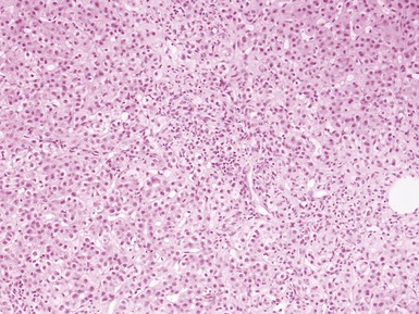
NEONATAL HEPATITIS
• The main histological differential diagnosis of prolonged neonatal cholestasis is neonatal hepatitis


ALPHA-1-ANTITRYPSIN DEFICIENCY


CYSTIC FIBROSIS


PROGRESSIVE FAMILIAL INTRAHEPATIC CHOLESTASIS (PFIC)
• Group of rare autosomally inherited disorders of transport of conjugated bile acids into the bile canaliculus


BILE ACID SYNTHESIS DEFECTS


LYMPHEDEMA–CHOLESTASIS SYNDROME
(Aagenaes syndrome; Drivdal et al 2006)