Fig. 6.1
(a) Longitudinal section, showing hypoechoic and intramural myoma in the posterior region of the cervix (red arrow), compressing the cervical canal. (b) Color Doppler showing central and peripheral vasculature in the myoma
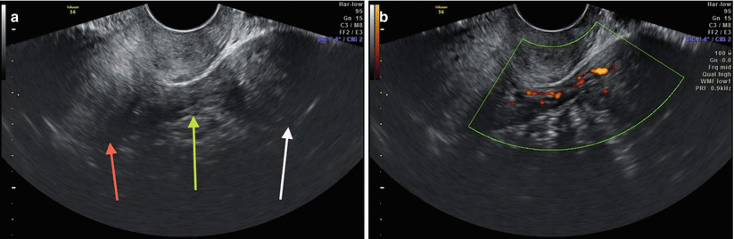
Fig. 6.2
(a) Axial ultrasound section, showing uterine body (red arrow), pedicle myoma in the left adnexal region (white arrow) and its respective pedicle (green arrow). (b) Power Doppler showing vascularization in vascular pedicle of the myoma

Fig. 6.3
(a) Axial ultrasound section of the uterine body (red arrow), showing subserosal nodule in the left side wall that compresses the proximal portion of the left uterine tube (green arrow). (b) Power Doppler showing sparse vascularity within the myoma

Fig. 6.4
(a) Axial ultrasound section of the uterine fibroid, showing myoma (red arrow) with submucosal component, less than 50%, and mostly with intramural component. The green arrow indicates the endometrial echo. (b) Longitudinal ultrasound section of the uterus, showing peripheral vascularization in the myoma, in a study with power Doppler. (c) Same myoma (red arrow) in longitudinal section, showing greater intramural than submucosal component. Note the narrowing of the anterior portion of the endometrium

Fig. 6.5
(a) Longitudinal ultrasonographic section of the uterus, showing, between the arrows, oval and hyperechoic focal thickening of the endometrial echo, compatible with polyp. (b) Power Doppler demonstrating vascular pedicle in the polyp

Fig. 6.6
Longitudinal section of retroverted uterus. The yellow arrow shows oval and hyperechoic image, in endometrial echo, compatible with polyp. Notice the previous endometrial bulging

Fig. 6.7
(a) Hysterosonography: Longitudinal ultrasound section of the uterus. The blue arrow shows a previous subserosal myoma. The yellow arrow shows anechoic fluid in the endometrial cavity (saline solution infused) that works as a contrast, allowing better identification of the endometrial polyp (red arrow). Notice the clear delineation between the endometrium and myometrium. (b) Same hysterosonography, showing the polyp in coronal section, in 3-D ultrasound, with measures between the calipers

Fig. 6.8
Coronal section, in 3-D ultrasound. The picture shows the endometrial cavity, with a small amount of liquid. The white arrows show two adjacent nodes, compatible with polyps. The good delimitation between the endometrium and the myometrium is evident

Fig. 6.9
(a, b) Asherman’s Syndrome (also known as “uterine synechiae”) is a condition characterized by the presence of scarring or fibrosis within the uterine cavity. In general, uterine synechiae are caused by damage to the lining of the uterus, such as curettage after abortion (most common), endometritis, intrauterine surgery, and radiotherapy; these can cause infertility, recurrent miscarriages, and menstrual disorders. The prognosis for success in pregnancy after the removal of uterine adhesions is associated with the type and severity of synechiae. Endometrial synechiae have excellent prognoses. On the other hand, the fibrous synechiae can only be undone with resection or laser, and have a high rate of recurrence after resection, making the prognosis of pregnancy much worse. The white arrows show the presence of thin, hypoechoic tissue “dividing” the endometrial echo (synechiae) in a 32-year-old patient with difficulty getting pregnant after curettage

Fig. 6.10
Hysterosonography, showing longitudinal section of the uterus. The yellow arrow indicates the catheter balloon used. The red arrow shows liquid infused into the endometrial cavity. The heads of white arrows show a large submucosal nodule (myoma). Note that − unlike the polyps − the endometrial interface with the myometrium is poorly defined due to the presence of the nodule

Fig. 6.11


MRI of the pelvis, sagittal, T2 weighted, showing uterine nodule with hyposignal (arrow), presenting intramural and submucosal component
Stay updated, free articles. Join our Telegram channel
Full access? Get Clinical Tree
