27 Juvenile Dermatomyositis
Juvenile dermatomyositis (JDMS) is the most common inflammatory myositis during childhood, accounting for more than 80% of cases. Endothelial injury of the capillaries, venules, and small arteries of the muscle, skin, and gastrointestinal tract characterize JDMS. It is a distinct disease from adult-onset dermatomyositis because it is not associated with malignancy and tends to remit after several years.
Etiology and Pathogenesis
The cause of JDMS is unclear; however, roles for genetics, environmental exposures, and infections have been postulated. Certain human leukocyte antigen (HLA) alleles are more common in patients with JDMS, including HLA-DQA1*0501 and HLA-DQA1*0301. Genetic polymorphisms in non–HLA-associated genes, such as tumor necrosis factor-α (TNF-α) and interleukin-1 receptor antagonist, have also been associated with JDMS. The presence of antinuclear antibodies (ANA) in 50% to 70% of patients provides some evidence for immune dysregulation. It has been suggested that viral infections, such as Coxsackie B or echovirus, may trigger an unusual immune response in a genetically susceptible host. However, this theory has yet to be substantiated.
Clinical Presentation
JDMS is a rare inflammatory myopathy with an incidence of two to four cases per million per year in children ages 16 years and younger. The mean age of onset is 7 years. However, there is a bimodal distribution with peaks at 2 to 5 years and 12 to 13 years. It is more common in girls than boys by a ratio of 2 : 1. There is no racial predominance.
Cutaneous Features
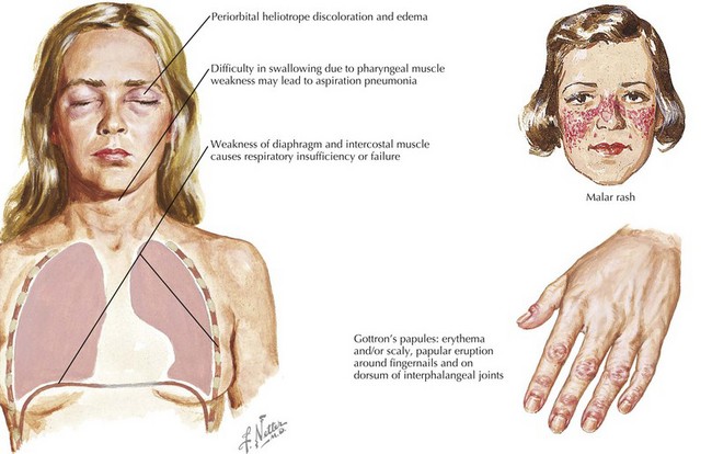
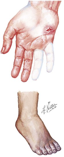
Dermatomyositis has several classic cutaneous findings (Figure 27-1). Gottron’s papules are erythematous, raised, scaling plaques on the dorsal surface of the knuckles, elbows, and knees that are present in more than 90% of children at the time of diagnosis. Gottron’s papules are frequently confused with severe eczema if JDMS is not suspected. The classic heliotrope rash is a violaceous discoloration of the eyelids with associated eyelid edema; this is present in approximately 80% of children at diagnosis. About 40% of children also have a prominent erythematous malar rash. This rash can be ulcerative, cross the nasolabial folds, and extend onto the forehead. Raynaud’s phenomenon (Figure 27-2) and associated nailfold capillary changes are seen in up to 80% of patients. Nailfold changes are characterized by proximal nailfold erythema, capillary dilatation, tortuosity, or dropout. The measured density of capillaries per millimeter may be a useful tool for monitoring clinical activity. Skin ulcerations reflect significant vasculopathy of the skin and may be a sign of internal organ vasculopathy. Ulcerative skin lesions are associated with more severe disease and worse prognosis.
Calcinosis, or calcium deposition in the skin and subcutaneous tissues, occurs in up to 40% of patients within a few years of diagnosis. Risk factors for the development of calcinosis include delayed diagnosis or treatment, TNF-α-308a genotype, and an age younger than 5 years at the time of diagnosis. Calcinosis may regress after disease remission, but complete resolution may take years. The calcinosis associated with JDMS occurs in five distinct patterns: (1) superficial calcinosis on the extremities that does not interfere with function and often regresses spontaneously; (2) deep tumoral muscle calcification in the proximal muscle groups that may interfere with joint motion, may ulcerate, and may require surgical debridement; (3) diffuse and painful calcinosis along myofascial planes that limits joint motion; (4) mixed forms of the former three types; and (5) extensive exoskeleton-like calcium deposits that result in severe functional limitations.
Other less common skin manifestations associated with JDMS are “mechanics’ hands” (thickening of the margins of the palms and radial surfaces of the hands), the shawl sign (macular erythema on the posterior neck and shoulders), the V sign (macular erythema on the anterior neck and chest), and poikiloderma vasculare atrophicans (circumscribed violaceous erythema with associated telangiectasia, hypopigmentation or hyperpigmentation, and superficial atrophy). The scalp may also be involved with seborrhea-like scaling, atrophy, and alopecia. Lipodystrophy is often underappreciated and may be present in 20% to 50% of children; it may be associated with insulin resistance, decreased glucose tolerance, acanthosis nigricans, and elevated triglycerides.

Stay updated, free articles. Join our Telegram channel

Full access? Get Clinical Tree

