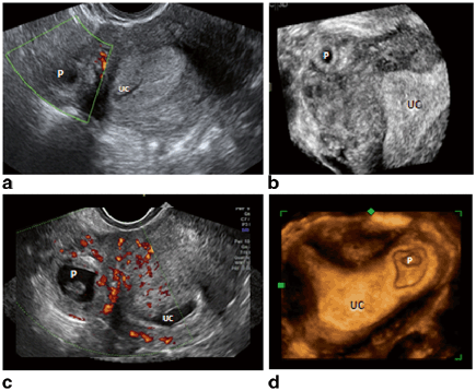
Fig. 11.1
Transvaginal ultrasound (TVUS). a An empty uterine cavity (UC) with echogenic line (arrows) leading towards gestational sac (P). b 3D TVUS rendering showed thin myometrial edge marked with calipers abutting gestational sac (P). Note the empty uterine cavity (UC). c TVUS revealed vascularity surrounding the gestation (P). The uterine cavity was empty (UC). d 3D TVUS tomographic rendering showed ectopic gestation denoted by P with empty uterine cavity (UC)
My Management
1.
Expectant Management
2.
Methotrexate administration
3.
Laparoscopic resection
Diagnosis and Assessment
Our patient demonstrates common presentation of interstitial pregnancy . Interstitial pregnancy implants in the segment of fallopian tube that traverses the muscular wall of the uterus. Its incidence is estimated to be 2–4 % of ectopic pregnancies and 1 in 2500–5000 live births. It is also known as “cornual pregnancy,” found in the outermost horn of the uterus, though this term should be reserved for patients with bicornuate uterus. Due to the thickened diameter of the outermost fallopian extension, there is a potential for expansion. Though rupture most commonly occurs prior to 12 weeks in 22 % of patients, expansion allows many interstitial pregnancies to remain asymptomatic for upwards of 7–16 weeks [1−3].Mortality for undiagnosed condition reaches 2.5 %, which is sevenfold higher than for other ectopic locations. This is most commonly due to catastrophic hemorrhage involving late rupture [4].
Assessment should be initiated with TVUS, which is the primary method for diagnosis; it is not only cost effective but also readily available. An empty uterine sac, paucity of myometrium around the superior-lateral portion of the gestational sac (GS), a distance of at least 1 cm from the edge of the uterine cavity to the GS, and an interstitial line are key identifiers for correct diagnosis [5−8]. As described by Ackerman, the interstitial line is an echogenic line that runs from the endometrial cavity to the cornual region, abutting the interstitial mass or GS. Identification of these diagnostic markers yields specificity for interstitial pregnancy of 88–98 % (Fig. 11.2).


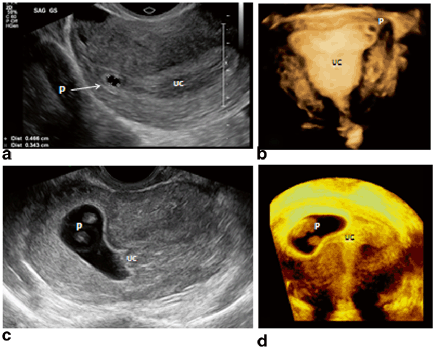
Fig. 11.2
TVUS images concerning ectopic gestation. a TVUS reveals empty uterine cavity. b 3D TVUS confirms interstitial pregnancy. c Note empty uterine cavity (UC). d 3D TVUS reveals intrauterine gestation (P) in UC at left horn. TVUS transvaginal ultrasound. (Images courtesy of Dr. Ilan Timor)
Three-dimensional (3D) ultrasound is especially useful in the diagnosis of interstitial pregnancy (Figs. 11.2 and 11.3). Navigating in the orthogonal planes in order to generate the desired section is helpful. Using the tomographic, multi-sectional display allows systematic scrutiny. Employing the thick slice rendering of volume contrast imaging (VCI) enables the investigator to achieve a particularly sharp edge enhancement in order to differentiate the hyperechoic, decidualized endometrium from intervening myometrium and the eccentrically located GS. A 3D TVUS should be the diagnostic tool of choice for accurate diagnosis [6, 8].


Fig. 11.3
TVUS images concerning for ectopic gestation. a TVUS reveals pregnancy (P) appears outside of UC. b 3D TVUS reveals intrauterine gestation in left horn. c TVUS concerning for empty UC. d 3D TVUS confirms interstitial pregnancy (P) with empty UC. Transvaginal ultrasound (TVUS), uterine cavity (UC). (Images courtesy of Dr. Ilan Timor)
When utilizing TVUS, color flow mapping should be employed to aid in identification of blood supply to the GS. In addition, the use of 3D ultrasonography, as demonstrated in Fig. 11.2 b, d and 11.3 b, d, as well as tomographic planning adds vital details of myometrial measure and exact pregnancy location.
If the rare event that 2D and 3D TVUS are inconclusive, MRI may be considered for diagnostic differentiation. Criteria are similar, including an eccentric GS, scant myometrial tissue surrounding the GS, and detection of the interstitial line [9].
Management
If an interstitial or cornual pregnancy is identified without the presence of fetal cardiac activity, expectant management is a viable option. However, once positive fetal heart rate is identified, the risks of rupture and subsequent maternal morbidity necessitate other interventions [8].
Stay updated, free articles. Join our Telegram channel

Full access? Get Clinical Tree


