(1)
Department Obstetrics & Gynaecology, St George Hospital, Kogarah, New South Wales, Australia
Abstract
Unlike the other conditions considered in this text which are very common, interstitial cystitis (IC) is quite rare. This chapter is a short summary of a great deal of research and clinical studies that have been directed at this fascinating problem. Appropriate textbooks are recommended.
Unlike the other conditions considered in this text which are very common, interstitial cystitis (IC) is quite rare. This chapter is a short summary of a great deal of research and clinical studies that have been directed at this fascinating problem. Appropriate textbooks are recommended.
How to Diagnose It
IC is a chronic pain syndrome characterized by the following:
Recurrent episodes of suprapubic pain/pelvic pain. Pain is worse when the bladder is full. The symptom of urgency is actually painful (in 92 % of cases).
Severe frequency (can void 60 times per day or more).
Generally, severe nocturia (ten times per night or more, in 51 % of cases).
Leakage of urine is not typical (but can occur).
Less common symptoms include the following:
Chronic pelvic pain or pressure symptoms (64–69 %)
Dysuria (61 %)
Dyspareunia (55 %)
Pain for days after sexual intercourse (37 %)
Hematuria (22 %)
IC is more common in women (ratio 9:1).
Large studies indicate prevalence of about 18 per 100,000 women Ho et al. [3]. The annual estimated incidence is 2.6 per 100,000 total US population. The average IC patient sees three or four urologists or gynecologists before diagnosis.
The diagnosis of IC is based on:
The classic symptoms of pain with frequency/urgency/nocturia. The Frequency Volume Chart (FVC) shows severe frequency/ nocturia. FVC usually shows small volumes/small bladder capacity. Urodynamic testing is painful and just shows small bladder capacity (although in some cases detrusor contractions are seen). Voiding function is usually normal (flow rate and residual urine). Urine cultures are generally sterile (by definition must be sterile for 3 months). Cystoscopy must be performed under general anesthesia (GA).
Mucosa often fairly normal during first fill.
Capacity under GA is reduced, for example, 400–600 ml.
Refill exam must be performed: shows petechial hemorrhages and small punctate red dots scattered over the mucosa.
In severe cases, may see Hunner’s ulcers—red splits or cracks in the mucosa.
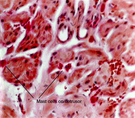
Bladder biopsy is recommended. Biopsy needs special stains for mast cells (see Fig. 12.1). It often but not always reveals excess mast cells in the detrusor muscle.


Figure 12.1
Mast cells in the detrusor muscle from a biopsy taken from patient with classic features of IC
Etiology
The etiology of IC remains unknown, although several theories have been put forward.
The Defective Epithelial Barrier Theory: The bladder mucosa is lined by a chemical layer of glycosaminoglycans (GAGs) which are thought to render the urothelium impermeable to harmful solutes (such as urea). Early histological studies suggested that the urothelium of IC patients was more readily penetrated, but later functional radioisotope studies showed no significant differences between IC patients and controls.
Stay updated, free articles. Join our Telegram channel

Full access? Get Clinical Tree


