Injury and Trauma

8 Injury and Trauma


Injury is one of the most important public health threats to children in the United States. Trauma causes more deaths in children and adolescents than all other causes combined. The care of injured children is a vitally important and specialized skill. Differences between child and adult anatomy and physiology make injury patterns and responses to injury unique to the pediatric age group.


Effective trauma resuscitation and treatment require the rapid acquisition and communication of large amounts of information about patients’ respiratory and cardiovascular stability as well as the extent of their injuries. It is common practice to follow a structured examination technique and management strategy during the evaluation of trauma patients, such as that taught in Advanced Trauma Life Support (ATLS) courses. Use of these principles helps to minimize secondary injury in pediatric trauma victims and ensures the best possible outcomes in these patients.


Etiology and Pathogenesis


Injury is the leading cause of death from ages 1 to 19 years in the United States. In addition, injury causes 30% of all deaths in children younger than 1 year of age. Eighty percent of all deaths related to trauma occur at the scene of the injury or in the emergency department (ED). This highlights the need for effective prevention strategies and the importance of appropriate prehospital and ED care for pediatric trauma victims.


The most common causes of injuries and resultant injury patterns are age specific. In infants (younger than 1 year of age), a high percentage of injuries are nonaccidental in nature, and it is important to maintain a high index of suspicion for possible inflicted injuries (see Chapter 12). Falls are the most common cause of injury in children from 1 to 14 years of age. Motor vehicle collisions and motor vehicle–pedestrian incidents are the most common causes of fatal injuries in children ages 1 to 18 years. Drowning (see Chapter 6) is the second most common cause of fatalities in younger children, and firearm-related deaths are the next most common cause in adolescents ages 15 to 18 years.


In children and adolescents presenting to trauma centers because of injury, a head injury is part of the presenting findings in 60% of patients. Different patterns of injury are evident in children of various ages and can often be predicted by the mechanism of injury and the size and age of the patient. For example, when struck by motor vehicles, younger and smaller children may primarily sustain head injuries. Older toddlers and young school-aged children may sustain injuries consistent with Waddell triad’s (closed head injury, intraabdominal injury, midshaft femur fracture), and adolescents may sustain primarily extremity injuries. The clinical presentation of pediatric trauma victims varies depending on the mechanism and severity of their injuries. Evaluation and management are aimed at assessing the nature and severity of the patient’s injuries while beginning stabilization and deciding on the needed course of therapy.


Evaluation and Management


Primary Survey (see Chapter 1)


The primary survey is structured to generate information vital to the immediate treatment of life-threatening issues. It is often referred to as the ABCs (airway, breathing, and circulation) but is better thought of as ABCDE (airway, breathing, circulation, disability, and exposure and environment), which is delineated below. Children who have sustained injuries are at risk of secondary injuries caused by hypoxia, hypercarbia, and poor perfusion. Recognition and correction of these conditions are the goals of this rapid portion of their assessment.


Airway


Initial evaluation of the airway is of utmost importance. A child who can phonate normally has a patent airway. In an unconscious patient, inspection for foreign body in the pharynx and for midface or mandibular injuries, which could compromise airway patency, should occur, as well as observation of obvious stridor or stertor with respiratory effort. Hoarseness or crepitus over the tracheal cartilage should raise concern for laryngeal fracture. After these areas have been examined, the physician should quickly move to secure an airway if necessary or move on to the assessment of ventilatory efficacy.


Readers are directed to Chapter 1 for complete information about airway evaluation and management. However, it is important to reiterate here the need for inline stabilization of the cervical spine (c-spine) during this assessment and treatment, especially if intubation is attempted. The collar should be opened or removed before direct laryngoscopy because it can impede successful visualization of the larynx. An assistant should hold the patient in the midclavicular line with the ears firmly between the lower arms to eliminate movement of the c-spine during intubation.


Breathing


Evaluation of effective ventilatory effort occurs after the airway evaluation. The chest wall excursion should be observed for symmetric chest rise. Visual inspection, including of the axillae and back, should identify open wounds. The clinicians should evaluate for symmetry and adequacy of breath sounds by auscultating the chest. Asymmetric chest rise or asymmetric breath sounds suggest the possibility of pneumothorax, hemothorax, flail chest, or an open chest wound.


Asymmetry in breath sounds may signify pneumothorax or hemothorax. This is important to note on the initial examination even when it appears clinically insignificant because a simple pneumothorax can be quickly converted to a tension pneumothorax when a patient is ventilated with positive pressure by bag–valve–mask (BVM) or endotracheal ventilation. Also, children are especially sensitive to the development of tension pneumothorax because of their relatively compliant and mobile mediastinum. Radiographs can be used to determine the presence and amount of air or blood in the pleural space. In a more clinically stable patient, a small simple pneumothorax may be treated noninvasively with inpatient monitoring, oxygen therapy, and serial chest radiography. Larger pneumothoraces or hemothoraces should be managed with chest tube drainage.


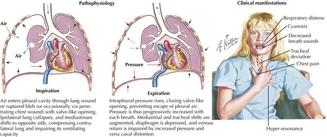
An absence of breath sounds, especially in conjunction with hypotension, should alert the clinician to possible tension pneumothorax. Tracheal deviation to the opposite side and hyperresonance of the chest to percussion may be less obvious in pediatric patients, and the more mobile mediastinum in this age group may produce vascular collapse in a more rapid fashion. Needle decompression can be immediately undertaken in such circumstances, with the introduction of a large-bore over-the-needle catheter (e.g., a 14-gauge intravenous [IV] catheter) into the second intercostal space in the midclavicular line. Needle decompression necessitates the subsequent placement of a chest tube. Massive hemothorax can present in a similar fashion, although the chest should be dull to percussion, and severe hypotension often predominates because of the significant blood loss (Figure 8-1).


Open chest wounds create loss of the usual negative pressure upon inspiration that is needed to inflate the lung. If the chest wall defect is large compared with the airway, air will preferentially be brought into the defect as opposed to the airway, significantly impairing effective ventilation. An occlusive dressing should be placed over sucking chest wounds, closed on three sides, to allow for air to escape during exhalation but preventing air from entering during inspiration.


Circulation


After the respiratory assessment, hemodynamic status should be evaluated by auscultation of heart sounds; palpation of central pulses (femoral or brachial), paying attention to both the rate and quality; and rapid evaluation of skin color, capillary refill, and level of consciousness. Any obvious area of external hemorrhage should be identified and addressed with pressure dressings; whip stitching; or in the case of massive scalp wounds, stapling to control bleeding.


Complete details on hypoperfusion and shock may be found in Chapter 2. In patients who have sustained significant traumatic injuries, IV access should be secured as quickly as possible with two large-bore IV catheters placed in large peripheral veins. If peripheral access attempts are unsuccessful in a patient showing signs of shock, clinicians should proceed quickly to obtain interosseous (IO) access. IO lines should not be placed distal to any obvious fractures.


Shock in a trauma patient should be presumed to be attributable to hypovolemia secondary to hemorrhage. Because most pediatric trauma victims have sustained blunt trauma, hemorrhage may be attributable to internal injuries such as splenic or hepatic lacerations and not readily visualized. However, other causes of traumatic shock must be considered, especially in patients with shock unresponsive to fluid resuscitation or with hemodynamic collapse. Chief among these causes are tension pneumothorax, tension hemothorax, and cardiac tamponade.


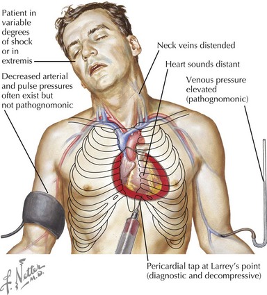
Cardiac tamponade is more common with penetrating chest wounds but can be caused by rupture of the myocardium with blunt traumatic force. As with tension pneumothorax, patients with cardiac tamponade present with tachycardia, hypotension, and distended jugular veins. However, with tamponade, there is no tracheal deviation. Pulsus paradoxus, which is exaggeration of the normal decrease in blood pressure with spontaneous inspiration, may be present in association with tamponade. Rapid bedside ultrasonography can help make the diagnosis, but if unavailable or if the patient is hemodynamically unstable, pericardiocentesis can be used to diagnose and treat tamponade (Figure 8-2).


Finally, hypotension in the pediatric trauma patient can, rarely, be caused by spinal cord injury. This should be suspected in a patient with an appropriate injury mechanism who is observed to have bradycardia, or a lack of appropriate tachycardia, in the face of hypotension.


The rapid installation of IV fluids is standard first-line therapy for shock. Boluses of 20 mL/kg of 0.9% saline solution or lactated Ringer’s solution should be given rapidly. If there is no response after 40 mL/kg is administered, then blood products should be given. In a child with decompensated shock caused by trauma, O-negative packed red blood cells (PRBCs) should be pushed rapidly until type-specific or crossmatched blood is available. In patients who require massive transfusion, fresh whole blood or fresh-frozen plasma and platelets in addition to PRBCs should be considered. If shock is not rapidly reversed with transfusion therapy, identification of the source of hemorrhage must be achieved, and operative management to halt the hemorrhage must be immediately undertaken.


Disability


After the patient’s hemodynamic status has been stabilized, the neurologic status is evaluated. The symmetry, size, and briskness of pupillary response should be documented along with the level of responsiveness. Numerical scales, such as the Glasgow Coma Scale (GCS, see Figure 1-8), are used to generate structured assessments of the level of responsiveness. This allows for comparisons in mental status evaluations over time, comparisons of assessments performed by different clinicians, and improved communication among providers. The GCS has been adapted for use in preverbal patients as well. A more straightforward, although somewhat less nuanced, system for rapid neurologic evaluation is the AVPU (awake, verbal, pain, unresponsive) system. Abnormalities in the pupillary response or level of consciousness should increase concern for intracranial injuries and alert the team to prepare for possible interventions such as intubation for airway protection or to improve ventilation, mannitol, or hypertonic saline administration for increased intracranial pressure (ICP) or emergent neurosurgical intervention.


In patients who are alert and can cooperate with the examination, the patient’s strength and sensation should be assessed in all four extremities. Tenderness and deformities of the cervical, thoracic, and lumbar spine should also be assessed. In preverbal children, this assessment can be challenging. Observation of spontaneous movement, watching how the patient reaches for objects, and the patient’s response to touch or painful stimulus should be assessed. The spine should be palpated, but it is difficult to ascertain tenderness in these children.


Secondary Survey


The goal of the secondary survey is to detail all likely injuries with a head-to-toe evaluation of the patient. It is best if this is done in an ordered way to avoid inadvertently leaving out body areas.


Stay updated, free articles. Join our Telegram channel

Jun 19, 2016 | Posted by in PEDIATRICS | Comments Off on Injury and Trauma

Full access? Get Clinical Tree

Get Clinical Tree app for offline access