Inflammatory Bowel Disease

110 Inflammatory Bowel Disease


Inflammatory bowel disease (IBD) is an idiopathic, likely immune-mediated, inflammatory disease involving the gastrointestinal (GI) tract. There are two subtypes, ulcerative colitis (UC) and Crohn’s disease. In the pediatric population, Crohn’s disease is more common than UC, with the majority of children diagnosed in adolescence, although some are much younger. Worldwide, the incidence of IBD is higher in developed countries than developing countries. Persons living in urban areas have increased rates of IBD compared with persons living in rural areas. Currently, there is no cure for IBD, and the goal of therapy in the pediatric population is maintenance of long-term remission and normal growth and nutritional status.


Clinical Presentation


The differential diagnosis for IBD is extensive and largely depends on the presenting symptoms (Table 110-1).


Crohn’s Disease


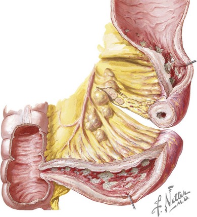
Crohn’s disease is characterized by transmural inflammation that may involve any segment of the intestinal tract from the mouth to the anus. Disease presentation is primarily determined by the location and extent of involvement. In children, the most common presenting distribution is ileocolonic disease (Figure 110-1) followed by small bowel disease alone and then colonic disease alone. Gastroduodenal disease is found in 30% of children with Crohn’s disease. Patients with ileocecal disease often present with right lower quadrant abdominal pain and diarrhea. In such patients, loops of bowel or fullness may be palpable in the right lower quadrant. Bloody stool is more common in colonic disease. Epigastric pain may occur with gastroduodenal disease. Dysphagia can be seen with esophageal involvement. Many children with Crohn’s disease have diarrhea, nocturnal defecation, low-grade intermittent fever, oral ulcers, weight loss, and decelerated growth velocity.


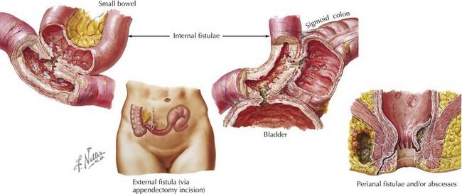
Crohn’s disease is categorized into three subtypes: inflammatory, stricturing, and fistulizing. Inflammatory disease is that described above. Stricturing disease involves luminal narrowing, which may be accompanied by prestenotic dilatation or signs of intestinal obstruction. This most commonly occurs in the small bowel but may involve the colon. Perianal or perirectal fistulae and abscesses or intraabdominal fistulae, phlegmon, or abscesses characterize fistulizing disease (Figure 110-2).


Extraintestinal Manifestations


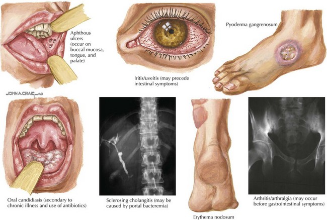
About a third of patients with IBD also have extraintestinal manifestations involving rheumatologic, cutaneous, ocular, vascular, hepatobiliary, renal, and skeletal systems (Figure 110-3). Arthralgia and arthritis are the most common extraintestinal symptoms and may involve both axial and peripheral joints. Joint manifestations may precede GI tract disease, so IBD should be in the differential diagnosis of isolated arthralgia or arthritis. Juvenile rheumatoid arthritis and ankylosing spondylitis are associated with IBD.


Skin manifestations of IBD are common in the pediatric population and include erythema nodosum and pyoderma gangrenosum. Red, raised, tender nodules located along the anterior shins characterize erythema nodosum. Its course correlates with the severity of GI tract inflammation. Deep necrotic ulcers (often on the legs) characterize pyoderma gangrenosum, with a course that is not related to intestinal disease.


The most common hepatobiliary diseases associated with IBD are primary sclerosing cholangitis and autoimmune hepatitis. Primary sclerosing cholangitis is more commonly associated with UC but can also be found in patients with Crohn’s disease. Its activity is not associated with intestinal disease activity, and its onset may occur years before or after IBD diagnosis. Other associated hepatobiliary disorders include hepatic abscess, hepatic granuloma, cholelithiasis, and cholecystitis. Cholelithiasis in IBD is caused by terminal ileal inflammation or resection, which leads to interrupted enterohepatic circulation of bile acids, causing lithogenic bile. Patients with ileal disease also have increased risk of kidney stones because of malabsorption of fat, which binds calcium and leaves oxalate to be absorbed and excreted in the urine.


Ocular findings associated with IBD include episcleritis, uveitis, and iritis. They are more common in Crohn’s disease patients compared with those with UC. Uveitis may be asymptomatic, so patients should have routine ophthalmologic examinations.


There is an increased incidence of thromboembolic disease in both Crohn’s disease and UC patients compared with the general population. This hypercoagulable state is likely attributable to chronic inflammation and may result in deep venous thrombosis, pulmonary embolism, and cerebrovascular disease.


IBD-associated osteopenia is multifactorial with contributions from malabsorption, malnutrition, inadequate calcium and vitamin D intake, chronic steroid use, inactivity, and chronic inflammation.

< div class='tao-gold-member'>

Stay updated, free articles. Join our Telegram channel

Jun 19, 2016 | Posted by in PEDIATRICS | Comments Off on Inflammatory Bowel Disease

Full access? Get Clinical Tree

Get Clinical Tree app for offline access