Infections of the Head and Neck

87 Infections of the Head and Neck


Cervical Lymphadenitis


Cervical lymph node enlargement is a common problem in children and adolescents. Almost all children have small palpable cervical lymph nodes. Cervical lymphadenopathy is defined by lymph nodes measuring more than 1 cm in diameter. Lymphadenitis refers specifically to inflammation of lymph nodes and is characterized by enlarged and tender nodes with warmth or erythema of the overlying skin.


Etiology and Pathogenesis


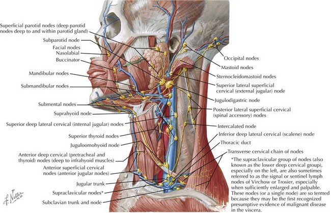
The cervical lymphatic system consists of a collection of both superficial and deep lymph nodes that protect the head, neck, nasopharynx, and oropharynx against infection (Figure 87-1). Lymph nodes can enlarge by either proliferation of normal cells intrinsic to the node such as lymphocytes or infiltration by cells extrinsic to the node such as neutrophils or malignant cells. The most common cause of cervical lymphadenopathy in children is reactive intranodal hyperplasia secondary to infection. The majority of lymphatics of the head and neck drain to the submandibular lymph nodes and the anterior and posterior cervical lymph node chains. Consequently, these nodes are involved in most children with cervical lymphadenitis.


Many different organisms have been implicated in cervical lymphadenitis (Box 87-1). The most common cause of cervical lymphadenitis is viruses that infect the upper respiratory tract, including adenovirus, respiratory syncytial virus, influenza, and parainfluenza. When bacterial in origin, cervical lymphadenitis may be a primary process or result from direct extension of a local infection such as pharyngitis or dental abscess. In the case of acutely inflamed and enlarged unilateral nodes, aspirates reveal infection by Staphylococcus aureus or Streptococcus pyogenes (group A β-hemolytic streptococci [GABHS]) in the majority of cases. Recent studies of suppurative lymphadenitis show the predominance of S. aureus and the increased prevalence of community-acquired methicillin-resistant S. aureus (CA-MRSA). More indolent causes of cervical lymphadenitis include Bartonella henselae, mycobacterial infections, and Toxoplasma gondii. The age of a child plays a role in predicting the infectious etiology of cervical lymphadenitis (Table 87-1).


Table 87-1 Etiology of Cervical Lymphadenitis by Age Group


Age Etiology
Infants Staphylococcus aureus
Group B streptococcus
Children age 1-4 y S. aureus
Group A streptococcus
Nontuberculous mycobacterium
Bartonella henselae
School-age children and adolescents S. aureus
Group A streptococcus
Anaerobes
B. henselae
Toxoplasma gondii


Clinical Presentation and Differential Diagnosis


The presentation of cervical lymphadenitis can be divided into three broad categories: (1) acute bilateral, (2) acute unilateral, and (3) subacute or chronic. The most common causes of acute bilateral cervical lymphadenitis are viral upper respiratory tract infections followed by pharyngitis caused by GABHS. In general, the lymph nodes are small, soft, and mobile without associated erythema, warmth, or significant tenderness. Additional clinical features such as gingivostomatitis in herpes simplex virus or pharyngoconjunctival fever caused by adenovirus may help to identify the causative virus. Viral causes of generalized lymphadenopathy, such as Epstein-Barr virus (EBV) and cytomegalovirus (CMV), can cause acute bilateral cervical lymphadenitis associated with infectious mononucleosis. In both cases, posterior cervical lymph node enlargement is most prominent followed by anterior cervical nodes.


Acute unilateral cervical lymphadenitis is caused by S. aureus and S. pyogenes in the majority of cases. The onset may be associated with an upper respiratory tract infection, pharyngitis, or periodontal disease, and associated fever is common. Typically, the onset is acute with development of large, tender, erythematous, and warm lymph nodes that may become fluctuant over a few days (Figure 87-2). In addition, a cellulitis–adenitis syndrome caused by group B streptococcus in infants between 3 and 7 weeks of age is associated with irritability, fever, and unilateral facial or submandibular swelling with erythema and tenderness.


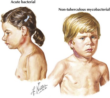
The most common causes of subacute or chronic lymphadenitis are mycobacterial infections, cat scratch disease, and toxoplasmosis. Lymph node enlargement is typically gradual in onset and progresses over weeks to months. The most common presentation of nontuberculous mycobacterium (NTM) disease in children is cervical lymphadenitis. The lymph nodes are large and indurated but nontender, and the overlying skin often becomes violaceous and thin (see Figure 87-2). Untreated lymphadenitis caused by NTM may resolve, but often it progresses to lymph node necrosis followed by fluctuance and spontaneous drainage. Cervical lymphadenitis caused by Mycobacterium tuberculosis has a similar presentation, but there are clinical and epidemiologic differences. NTM is uncommon in children older than 5 years of age compared with tuberculosis, which can occur at any age. With NTM, involvement is usually unilateral and associated with a normal chest radiograph and a normal or minimally indurated purified protein derivative (PPD). In contrast, children with tuberculous cervical lymphadenitis are more likely to have bilateral lymph node involvement, systemic symptoms, an abnormal chest radiograph, and an abnormal PPD result.


Cat-scratch disease, caused by B. henselae, most commonly affects the axilla and cervical regions. Most patients have a history of recent contact with cats or kittens. Clinical manifestations begin with a papule or pustule that develops at the inoculation site a few days to weeks after a bite or scratch followed by lymphadenitis proximal to the site. Lymphadenitis is tender and erythematous and often associated with fever. Lymphadenitis typically persists for several weeks to months and may suppurate. Acquired Toxoplasma infection, when symptomatic, generally presents as cervical lymphadenopathy and fatigue without fever. Lymphadenitis most frequently involves a solitary node in the head and neck region without systemic symptoms. Lymphadenitis secondary to toxoplasmosis tends to be nonsuppurative and may persist for many months. In addition, viral etiologies such as EBV, CMV, and HIV can cause bilateral subacute cervical lymphadenitis.


Noninfectious causes of lymphadenitis in children are less common but should always be considered in the differential diagnosis. Congenital cysts such as branchial cleft cysts, cystic hygromas, and thyroglossal duct cysts can mimic lymphadenitis, especially when infected. Malignancies such as lymphoma, leukemia, neuroblastoma, and rhabdomyosarcoma can present as cervical lymphadenopathy. Malignancy should be considered in cases of indolent lymphadenopathy, especially with a history of weight loss, fevers, night sweats, or lymphadenitis that is unresponsive to antibiotic treatment. Other causes of cervical neck masses should be included in the differential diagnosis of cervical lymphadenitis (Box 87-2).


Evaluation and Management


Lymphadenitis is a clinical diagnosis based on physical examination findings of enlarged and inflamed palpable lymph nodes. The evaluation and management of cervical lymphadenitis is directed by a thorough history and physical examination. Patients with acute small, bilateral nodes with minimal tenderness along with symptoms of fever or respiratory tract infection most likely have a viral syndrome and can be managed conservatively with observation and supportive care. If GABHS pharyngitis is suspected, a rapid streptococcal antigen test or throat culture should be performed. In patients with acute large, unilateral, erythematous, and tender nodes associated with fever, bacterial infection is most likely. Antimicrobial therapy should be directed at S. pyogenes and S. aureus with cephalexin being a reasonable choice. Because of the continuing rise of CA-MRSA in many areas, clindamycin is also an appropriate first-line option. In older children and adolescents, anaerobic bacteria should be suspected in cases of cervical lymphadenitis associated with gingival infections or dental abscesses. In these cases, amoxicillin–clavulanate or clindamycin provide anaerobic coverage in addition to gram-positive coverage. Cervical lymphadenitis can be managed on an outpatient basis for most children who are well-appearing, well-hydrated, and have no evidence of abscess. Hospital admission for intravenous (IV) antibiotics should be considered in infants, ill-appearing children, children who have fluctuant nodes or associated cellulitis, and patients who have failed outpatient treatment. IV antibiotic choices include cefazolin, oxacillin, and ampicillin-sulbactam. In regions with a high prevalence of CA-MRSA, clindamycin or vancomycin provides adequate coverage. The total treatment course should be 10 to 14 days. If there is no response to antibiotic treatment within 48 to 72 hours or clinical worsening, the next steps include evaluation for abscess formation. Ultrasound is useful to detect the presence and extent of an abscess if lymph node fluctuance is not obvious by examination. Surgeons may request a computed tomography (CT) scan before performing an incision and drainage to obtain more detailed imaging of adjacent and deep structures that may not be seen on ultrasound. However, ultrasound offers the advantage of avoiding radiation exposure. Aspirated or drained material from a suppurative node should be sent for Gram stain and both aerobic and anaerobic bacterial culture. Acid-fast stain and culture for mycobacterium and fungi should be considered with the appropriate clinical picture.


In cases of subacute or chronic lymphadenitis, evaluation for tuberculosis, NTM, HIV, EBV, CMV, cat scratch disease, and toxoplasmosis should be considered based on history and physical examination findings. A PPD can be helpful in distinguishing tuberculous from NTM lymphadenitis. Serologic testing is available for B. henselae, T. gondii, EBV, and CMV. Polymerase chain reaction is the preferred diagnostic test for HIV. Toxoplasmosis and cat scratch disease are typically self-limited infections that do not require treatment. For patients with systemic disease secondary to B. henselae, treatment with azithromycin is an option. However, the role of antimicrobial therapy in cat scratch disease remains controversial. With NTM lymphadenitis, excisional biopsy of the node can provide a definitive diagnosis and is the preferred treatment. If surgery is not feasible because of the location of involved nodes, clarithromycin or azithromycin with ethambutol or rifampin should be considered. When the etiology of cervical lymphadenitis remains unclear after negative infectious workup results, patients should be monitored closely for systemic signs of disease. An excisional biopsy may be necessary to establish the diagnosis in cases of persistent lymphadenitis that do not respond to appropriate antibiotic treatment or when nodes are nontender and fixed to adjacent tissue suggestive of malignancy.

< div class='tao-gold-member'>

Stay updated, free articles. Join our Telegram channel

Jun 19, 2016 | Posted by in PEDIATRICS | Comments Off on Infections of the Head and Neck

Full access? Get Clinical Tree

Get Clinical Tree app for offline access