Fig. 9.1
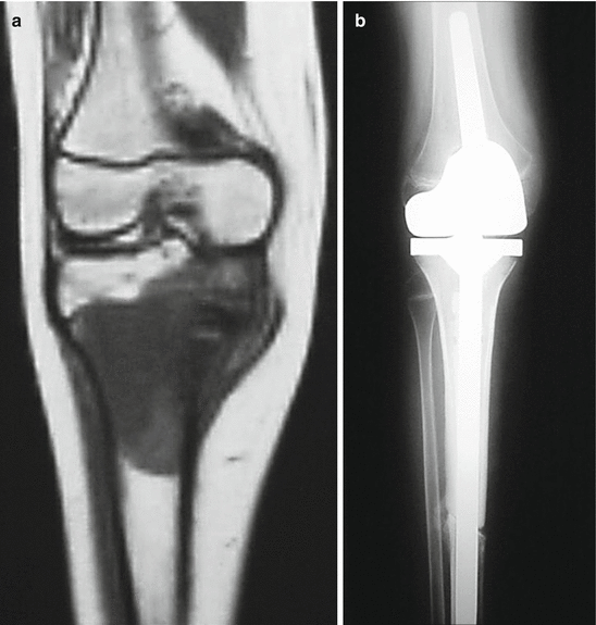
(a) Osteosarcoma in the distal metaphysis of the femur of a 15-year-old boy. The tumor seems to have crossed the physis in the MRI image. (b, c) However, the histological study found no tumor cells in the physis
9.2 Stages of Invasion of the Epiphysis
Several years ago, in our department, we carried out a retrospective histological study of a series of malignant bone tumors in children [2] (see Chap. 6). The proportion of cases in which the tumor infringed the physis, about 50 %, was similar to that found with our subsequent study of imaging methods (see Chap. 7). In the histological study, we found that morphological lesions at the physis could be categorized into three types:
The growth plate was not in contact with tumoral tissue.
Areas of the growth plate were in contact with tumor tissue but were not penetrated by the tumor. Voluminous capillary sinusoids had introduced themselves between the columns of the matrix of the cartilage. The remainder of the physis appeared to be free of alterations.
The physis was clearly invaded by the tumor. The areas crossed by tumor were surrounded by zones of thinned cartilage, similar to what was observed in the second type of lesion.
The implication of these observations is that invasion of the epiphysis by the tumor progresses in a predictable manner: first there is a hypervascularization reaction which leads to an early ossification of the growth plate, and after that the tumor crosses the physis.
9.3 Surgical Treatments
The surgical treatment we recommend for these tumors depends on the stage of invasion of the epiphysis as revealed by MRI. The three possibilities are presented below.
Get Clinical Tree app for offline access

1.
The tumor has crossed the physis.
In such cases, preservation of the epiphysis is not possible (Fig. 9.2).


Fig. 9.2
(a) The growth plate has been crossed by this osteosarcoma in the proximal metaphysis of the tibia. (b) Reconstruction with a composite allograft-prosthesis
2.
The tumor is in contact with the physis.
There are three scenarios:
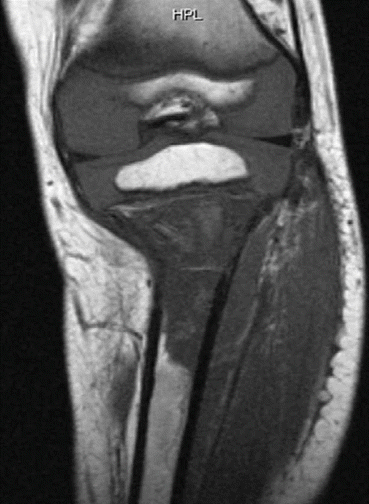
If all of the physis is affected (Fig. 9.3), the probability that tumor cells have crossed over the physis is high (see Chap. 5). However, most Ewing sarcomas and many osteosarcomas respond well to neoadjuvant chemotherapy, and if this response is particularly strong, preservation of the epiphysis should not be ruled out.

Fig. 9.3
Osteosarcoma in contact with the whole of the physis
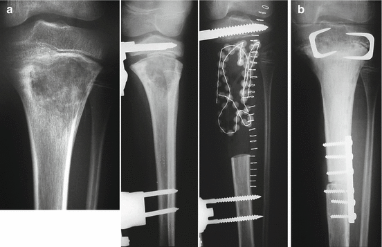
If the tumor is only in contact with part of the growth plate, tumor cells are less likely to have crossed over the physis and consequently we can try to preserve the epiphysis. After resection, external fixation can be maintained until intraoperative histological studies determine whether tumor cells are present in the physeal margin of the resection (Fig. 9.4). Then, on the basis of the histology, the appropriate manner in which to complete the surgical treatment (see Chap. 10) can decided. Before the advent of MRI, due to the lower accuracy of the other imaging methods employed, we used this methodology more frequently.

Fig. 9.4
Physeal distraction according to Cañadell’s technique in a case in which involvement of the physis was uncertain, before the MRI era. (a) External fixation was kept in place after resection until histological study of the resection margins had been carried out. (b) After histological confirmation of the absence of tumor cells in the metaphyseal margin of the resection, the reconstruction was carried out with an intercalary allograft
An alternative method for preserving the epiphysis in cases when a tumor is in contact with only a part of the physis but does not cross it is intra-epiphyseal osteotomy, which may be useful especially in certain cases in children who are nearing the end of growth.
3.
The tumor is near to but not in contact with the physis.
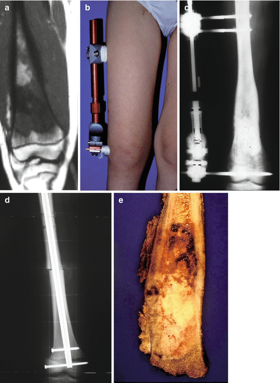
Physeal distraction before excision is, in our experience, the best technique in such cases (Fig. 9.5). The safety of physeal distraction and the fact that it can preserve the whole epiphysis and most of the growth plate make it superior to other techniques such as epiphyseal osteotomy.


