
Fig. 8.1
The communication skills with children at different ages. (a) Examination of infants: play with toys together to increase trust. (b) Examination of a young child aged 1–3: reward the child with candies to enhance his/her cooperation. (c) Examination of a young child aged 3–6: by means of verbal praise, lead the child through the examination in a light-hearted, happy manner. (d) Examination of a child aged 6–12: given a gentle verbal praise, most child patients can be actively cooperative for examination
8.1.2 Preschool Age (3–6 Years)
At this stage, the development of children’s brain function is reaching a plateau. Children exhibit an increase in vocabulary, a rapid progress in body movements, an expansion of living space, and a keen interest in all surroundings. Features in this period include (1) emotional instability and vulnerability to environmental influence; (2) showing talents for imitation and fondness of imitating their parents or actions of other child patients when being examined; (3) being playful, animated, and active; and (4) individuality beginning to take shape with a certain aptitude of self-control. Hence, pediatric ophthalmologists can well create a relaxing environment full of childishness and playfulness in the examination room. Moreover, they might as well ask parents and their children to do “role-playing” of seeing a doctor, give praise and award to the child patient who is cooperative for examination, and try to minimize the use of body immobilization and auxiliary sedatives (Fig. 8.1b, c).
8.1.3 School Age (6–12 Years)
At this stage, children’s capacities in comprehension, synthesis, analysis, and induction develop rapidly due to their intellectual maturity. A certain amount of self-discipline and tolerance has also developed. According to previous clinical statistics, roughly 95 % of child patients of school age are cooperative enough to be examined [3]. Examiners need to communicate patiently with them in a gentle voice, adopting the method of demonstration to get them actively involved in the examination. It is necessary for the examiner to be gentle in manipulation and try to shorten the duration of examination (Fig. 8.1d). Besides, the examiner can give proper praise to the child patient in order to stimulate his/her performance desire so as to facilitate examination.
8.2 History Taking
A detailed medical history must include a child patient’s basic information, chief complaint (CC), history of present illness (HPI), maternal pregnancy-labor history, the child’s birth, and family history.
Basic information: name, gender, age, date of birth, height, and weight.
Common chief complaint (CC): the child patient was found to have “white spots” in his/her eyes, predisposed to falling and getting hurt, with nystagmus, strabismus, squinting, short visual distance to an object, rubbing eyes with hands, photophobia, and binocular asymmetry. They could also be referred to the clinic after physical examination in the kindergarten and eye injury or when lens opacity is detected by other physicians. An ophthalmologist’s inquiries may begin with some simple questions (like patient’s visual acuity, whether he/she can follow light, whether he/she is cross-eyed when looking at objects, whether he/she often falls and gets hurt, whether he/she sits very close when watching television, and when he/she noticed the vision change) to know more about the child’s visual acuity. Due to the child’s short attention span and unwillingness to stay in unfamiliar surroundings, ophthalmologists should finish all the examinations rapidly after the child’s chief complaint is certain and then complete the history taking.
History of present illness (HPI): a detailed record of ocular abnormalities of the child patient, when they occurred, whether they had any causes or inducing factors, the progressions of the symptoms or any associated symptoms exist, and the examinations and the treatments given previously, including medication therapy or surgery.
Past medical history (PMH): a record of previous eye examination, drug allergies, glucocorticoid therapy (especially in the case of posterior subcapsular cataract), injuries, and surgeries.
Maternal pregnancy-labor history: maternal age at pregnancy, presence of any pregnancy-related diseases or infections during pregnancy (particularly TORCH infections, viz., toxoplasmosis, rubella virus, cytomegalovirus, herpes simplex virus 1 and 2, measles virus, and Treponema pallidum infection), presence of rash or fever during pregnancy (possibly suggesting recessive intrauterine infection), and presence of antenatal or perinatal factors (such as alcoholism, smoking, medications, and exposure to ionizing radiation during pregnancy).
Child’s birth: timing of birth (full term or premature), way of labor (eutocia, cesarean section, or other means of midwifery), and whether there was asphyxia, oxygen therapy, or incubation.
Family history of genetic diseases: About one third of children with congenital cataracts have family histories. Ophthalmologists should ask about the treatments and prognoses of the other patients within the families during history taking, which can serve as a reference for children’s postoperative visual function assessment [4].
8.3 Specialized Examinations of Pediatric Cataracts
Specialized examinations of pediatric cataract consist of evaluations of visual function and anatomical structures of the eye. However, child patients fail to cooperate with the examination, so anesthesia is usually needed.
8.3.1 Examination Under Anesthesia (EUA)
For a long time, specialized examination of pediatric eye diseases has been a tough issue for ophthalmologists across the globe. First of all, child patients are not cooperative enough to be examined. In addition, most of the ophthalmic examination equipments in practice are desktop machines that require examinees to remain seated. Although some handheld, noncontact devices have been recently developed, such as handheld slit lamp and handheld fundus camera, devices for adults are still used in the majority of pediatric eye examinations. All of these have caused inconvenience to the examinations, data analyses, and diagnoses of childhood eye diseases.
To resolve this tough issue, we have set up a series of effective methods of pediatric examination under anesthesia (EUA) aimed at various ages through years of clinical practice and experience.
8.3.1.1 Under Age 1 (Flying Baby)
During the examination, parents should work closely with the examiner. One parent or examiner lifts up the baby under the armpits with both hands and thumbs on the baby’s neck for support (Fig. 8.2a). The baby is held in a flying position with their head tilted forward to the forehead strap of the slit lamp (Fig. 8.2b). Another examiner or parent can put their left hand on the child’s occipital and help them get closer to the forehead strap. The right hand can also be put between the chin rest of slit lamp and the baby’s chin to stabilize the child’s head and protect it from collision. Then another examiner gently holds the child’s eyelids open, and the examination of the baby begins. The flying baby position facilitates smooth examination and manipulation.
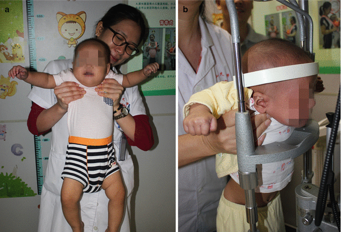
Fig. 8.2
Slit-lamp examination for a baby under age 1 year. (a) The infant is lifted up by the examiner in a flying position. (b) The examiner holds the infant in a flying position for slit-lamp examination
8.3.1.2 Age 1–3 Years (Baby Carrier)
As the baby has been gaining weight steadily, it is difficult for the parents to keep the baby “flying” for long. In this scenario, the examination can be performed with the help of a baby carrier. One parent holds the baby on the chest with the baby carrier and places the baby’s head close to the forehead strap with both hands (Fig. 8.3). The other parent lays his/her right hand over the chin rest to fix and protect the baby’s head, with the left hand in the baby’s occipital region, and gently tilts the baby’s head closer to the forehead strap. Lastly, the examiner gently holds open the baby’s eyelids and completes the examination and manipulation.

Fig. 8.3
Slit-lamp examination for a baby aged 1–3 years. (a) Mother holding her baby with a baby carrier. (b) Examiner helping the baby’s parent with slit-lamp examination using a baby carrier
8.3.1.3 Age 3 Years or Older (Flexible Bed)
Due to the baby’s weight and height, holding them or using a baby carrier alone does not ensure complete examination. In this case, a flexible bed for ophthalmic examination of children may be used (Fig. 8.4). Developed independently by the Home for Cataract Children, Zhongshan Ophthalmic Center (ZOC) of Sun Yat-sen University, the flexible bed for ophthalmic examination of infants and children possesses the functions of anesthesia bed and trolley. It has been patented in both China and the USA (China Patent No. ZL 201120251679.3; US Patent No. US9,015,882 B2). As a trolley, it can be used for the examination of child patients under anesthesia or in a conscious state. The height of the baby’s seat may be adjusted swiftly and freely as required by various table-mounted ophthalmic devices. During the examination, the parent may only need to push the flexible bed up to the slit lamp, with the right hand over the chin rest to stabilize and protect the baby’s head and the left hand in the baby’s occipital region to gently tilt their head closer to the forehead band. In doing so, the examiner can complete the examination smoothly and quickly.


