61 Hematuria and Proteinuria
Hematuria
Hematuria is the medical term for the presence of blood in the urine and is a common pediatric problem. Gross hematuria is clearly visible. Whereas blood cells of a glomerular origin are usually present in brown, tea-colored, or cola-colored urine, blood from the lower urinary tract changes urine to pink or red. Microscopic hematuria is defined by the presence of five or more red blood cells (RBCs) per high-power field on at least three occasions over a 3-week period in a spun urine sample. Out of every 1000 children presenting to an emergency department, nearly 1.5 have gross hematuria; 1% to 2% of school-aged children have microscopic hematuria.
Several substances besides blood can cause discolored urine. Microhematuria is generally first detected on a dipstick test but should be confirmed by microscopic examination of the sediment of spun urine. Gross hematuria, without casts, should always be evaluated by renal ultrasound.
All children should have a screening urinalysis at school entry (age, 4-5 years) and during adolescence (age, 11-21 years). Serious conditions should be considered; however, most children with isolated microhematuria do not have a severe illness and do not require extensive investigation.
Etiology and Pathogenesis
Table 61-1 and Box 61-1 provide a comprehensive differential diagnoses list for hematuria.
Table 61-1 Distinguishing Features of Glomerular and Nonglomerular Hematuria
Feature | Glomerular | Nonglomerular |
---|---|---|
History | ||
Burning on micturition | No | Urethritis, cystitis |
Systemic complaints | Edema, fever, pharyngitis, rash, arthralgias | Fever with UTIs; pain with calculi |
Family history | Deafness in Alport syndrome, renal failure | Usually negative except with calculi |
Physical Examination | ||
Hypertension | Often | Unlikely |
Edema | Sometimes present | No |
Abdominal mass | No | Wilms’ tumor, polycystic kidneys |
Rash, arthritis | SLE, HSP | No |
Urine analysis | ||
Color | Brown, tea or cola colored | Bright red or pink |
Proteinuria | Often | No |
Dysmorphic RBCs | Yes | No |
RBC casts | Yes | No |
Crystals | No | May be informative |
HSP, Henoch-Schönlein purpura; RBC, red blood cell; SLE, systemic lupus erythematosus; UTI, urinary tract infection.
Box 61-1 Differential Diagnosis of Hematuria
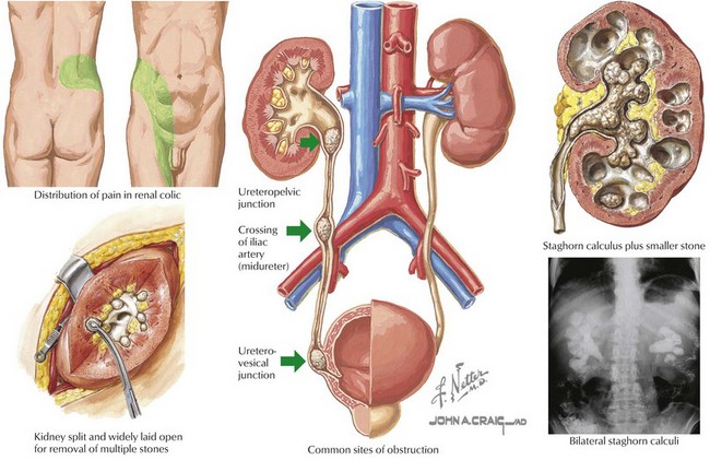
Hypercalciuria and Urolithiasis
Hypercalciuria can occur with low, normal, or high serum calcium levels. In eucalcemic patients, the most common cause is idiopathic; other causes include immobilization, Cushing’s syndrome, distal renal tubular acidosis, and Bartter’s syndrome. Disorders associated with hypercalcemia include hyperparathyroidism, vitamin D intoxication, hypophosphatasia, tumors, and immobilization bone resorption. Rare familial abnormalities of renal calcium channels are the cause of hypercalciuria with hypocalcemia. Hypercalciuria is the most common cause of stone disease.
Idiopathic hypercalciuria typically presents with asymptomatic microscopic hematuria. Stones can reveal their presence in a variety of ways to include dysuria, gross hematuria. and renal colic. Renal ultrasound or spiral computed tomography (CT) should be considered. Stones may be found incidentally after imaging is done for other reasons (e.g., radiographs for constipation). Radiographs can detect radiopaque stones but not those formed by uric acid.
The initial screening test for hypercalciuria is a urine calcium-to-creatinine ratio on a random urine sample. A ratio of greater than 0.2 in older children and adults is highly suggestive of hypercalciuria, with higher values in infants and young children. Confirmation should be obtained by a 24-hour urine collection with an excretion of greater than 4 mg/kg/24 hours. Urine from this collection should be tested for cystine, citrate, oxalate, phosphorus, and uric acid excretion as well. The collection is difficult in younger children, however. Serum chemistries, including calcium, phosphorus, magnesium, uric acid, urine pH, and renal function, should also be obtained. A detailed family history for stone disease, diet history, and evaluation of medications and nutritional supplements should be sought. Recovery of a stone should be attempted for analysis. Stones caused by infection are rare in children.
Management of these patients is twofold (Figure 61-1). Pain management should be optimized. Surgical intervention or lithotripsy is indicated in cases of urinary obstruction or recurrent stones with superimposed urinary tract infections (UTIs). After the cause has been determined, therapy to prevent stone recurrence can be implemented; this includes increased fluid intake to ensure dilute hypotonic urine, dietary manipulation, and drug therapy in some cases.
Medications
Microscopic hematuria caused by drug exposure is not uncommon. Although some medications are commonly used in children, most are not. Gross hematuria associated with cyclophosphamide can be severe in some cases.
Trauma
Traumatic injury to the urogenital tract is frequently seen with blunt force trauma and may be life threatening, depending on its severity. The presence of hematuria with minimal traumatic injury is highly suggestive of anatomic abnormalities of the kidney such as polycystic kidney disease.
Hematuria associated with renal trauma requires evaluation by CT scan, magnetic resonance imaging, or renal ultrasound. Urologic evaluation should be obtained before placement of a urethral or bladder catheter in cases of lower urinary tract trauma.
Most renal contusions or lacerations can be managed conservatively; however, significant lacerations of the kidney or injury to the collecting system or lower urinary tract may require emergent surgical intervention.
Malignancy
Wilms’ tumor is the most common childhood malignancy of the kidney and is discussed further in Chapter 53. Microscopic hematuria is more commonly found than grossly bloody urine, but hematuria as the presenting sign of Wilms’ tumor is rare.
Hematologic Causes
Hematuria commonly occurs with sickle cell hemoglobinopathy. Affecting patients with either sickle cell disease or sickle cell trait, it is thought to be caused by sickling and sludging of RBCs in the renal medulla. Typically painless and occurring in adolescent boys, it can be precipitated by trauma, exercise, dehydration, or infection. Papillary necrosis is also seen with severe dehydration and renal infarction.
Hematuria is rarely the sole finding in a patient with a coagulopathy. However, coagulopathies should be investigated in patients without another source for painless, gross hematuria and a history of bruising/bleeding, or a family history of a bleeding diathesis.

Stay updated, free articles. Join our Telegram channel

Full access? Get Clinical Tree

