Chapter 42 Gestational Trophoblastic Neoplasia
Gestational trophoblastic neoplasia (GTN) represents a unique spectrum of diseases that includes benign hydatidiform mole; invasive mole (chorioadenoma destruens), which can metastasize; and the frankly malignant variety, choriocarcinoma. Most molar pregnancies are sporadic, but a familial syndrome of recurrent hydatidiform mole has been described. Future research should lead to identification of the genetic defect responsible for this uncommon syndrome.
Most patients (80% to 90%) with GTN follow a benign course, with their disease remitting spontaneously. Most patients with metastatic disease can be effectively cured with chemotherapy. This diverse group of diseases has a sensitive tumor marker, human chorionic gonadotropin (hCG), which is secreted by all these tumors and allows accurate follow-up and assessment of the disease.
Epidemiology and Etiology
The incidence of molar pregnancy is about 1 in every 1500 to 2000 pregnancies among whites in the United States. There is a much higher incidence among Asian women in the United States (1 in 800) and an even higher incidence among women in Asia, for example, Taiwan (1 in every 125 to 200 pregnancies). The risk for the development of a second molar pregnancy is 1% to 3%, or as much as 40 times greater than the risk for developing the first molar pregnancy.
Although the cause of GTN is unknown, it is known to occur more frequently in women younger than 20 years and in those older than 40 years. It appears that GTN may result from defective fertilization, a process that is more common in both younger and older individuals. Diet may play a causative role. The incidence of molar pregnancy has been noted to be higher in geographic areas where people consume less β-carotene (a retinoid) and folic acid.
Genetics of Gestational Trophoblastic Disease
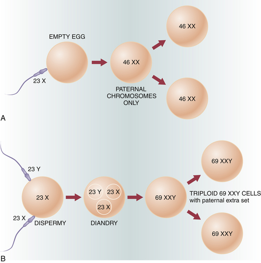
The cytogenetic analysis of tissue obtained from molar pregnancies offers some clue to the genesis of these lesions. Figure 42-1 illustrates the genetic composition of molar pregnancies.

FIGURE 42-1 Cytogenetic makeup of hydatidiform mole. A: Chromosomal origin of a complete mole. A single sperm fertilizes an “empty egg.” Reduplication of its 23 X set gives a completely homozygous diploid genome of 46 XX. A similar result follows fertilization of an empty egg by two sperms with two independently drawn sets of 23 X or 23 Y; note that both karyotypes, 46 XX and 46 XY, can ensue. B: Chromosomal origin of the triploid, partial mole. A normal egg with a 23 X haploid set is fertilized by two sperms that can carry either sex chromosome to give a total of 69 chromosomes with a sex chromosome configuration of XXY, XXX, or XYY. A similar result can be obtained by fertilization with a sperm carrying the unreduced paternal genome 46 XY (resulting sex complement, XXY only).
(Adapted from Szulman AE: Syndromes of hydatidiform moles: Partial vs. complete. J Reprod Med 29:789-790, 1984.)
COMPLETE MOLE
Most hydatidiform moles are “complete” moles and have a 46 XX karyotype. Specialized studies indicate that both of the X chromosomes are paternally derived. This androgenic origin probably results from fertilization of an “empty egg” (i.e., an egg without chromosomes) by a haploid sperm (23 X), which then duplicates to restore the diploid chromosomal complement (46 XX). Only a small percentage of lesions are 46 XY. Complete molar pregnancy is only rarely associated with a fetus, and this may represent a form of twinning.
PARTIAL MOLE
In the “incomplete” or partial mole, the karyotype is usually a triploid, often 69 XXY (80%). Most of the remaining lesions are 69 XXX or 69 XYY. Occasionally mosaic patterns occur. These lesions, unlike complete moles, often present with a coexistent fetus. The fetus usually has a triploid karyotype and is defective.
Classification
The term gestational trophoblastic neoplasia is of clinical value because often the diagnosis is made and therapy instituted without definitive knowledge of the precise histologic pattern. GTN may be benign or malignant and nonmetastatic or metastatic (Box 42-1).
The benign form of GTN is called hydatidiform mole. Although this entity is usually confined to the uterine cavity, trophoblastic tissue can occasionally embolize to the lungs. The malignant forms of GTN are invasive mole and choriocarcinoma. Invasive mole is usually a locally invasive lesion, although it can be associated with metastases. This lesion accounts for most patients who have persistent hCG titers following molar evacuation. Choriocarcinoma is the frankly malignant form of GTN.
Metastatic GTN can be subdivided into good prognosis and poor prognosis groups, depending on the sites of metastases and other clinical variables (Box 42-2).
Pathologic Features
Grossly, a hydatidiform mole appears as multiple vesicles that have been classically described as a “bunch of grapes” (Figure 42-2). The characteristic histopathologic findings associated with a complete molar pregnancy are (1) hydropic villi, (2) absence of fetal blood vessels, and (3) hyperplasia of trophoblastic tissue (Figure 42-3). Invasive mole differs from hydatidiform mole only in its propensity to invade locally and to metastasize.

FIGURE 42-2 Complete hydatidiform mole. Multiple hydropic villi (vesicles), resembling a bunch of grapes, are admixed with areas of necrosis (white areas) and hemorrhage. Note the absence of a fetus.

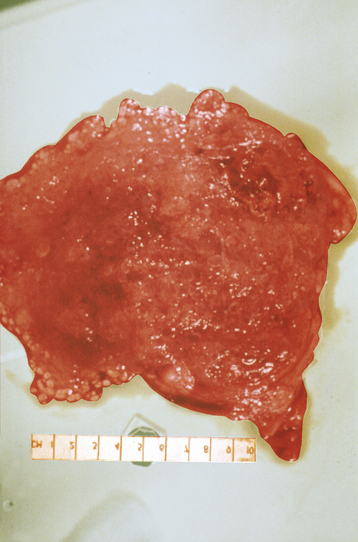
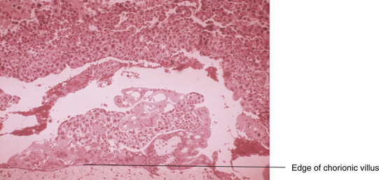
FIGURE 42-3 Histologic appearance of a complete hydatidiform mole. Note the marked trophoblastic proliferation.
A partial mole has some hydropic villi, whereas other villi are essentially normal. Fetal vessels are seen in a partial mole, and the trophoblastic tissue exhibits less striking hyperplasia.
Choriocarcinoma in the uterus appears grossly as a vascular-appearing, irregular, and “beefy” tumor, often growing through the uterine wall. Metastatic lesions appear hemorrhagic and have the consistency of currant jelly. Histologically, choriocarcinoma consists of sheets of malignant cytotrophoblast and syncytiotrophoblast with no identifiable villi.
Hydatidiform Mole
SYMPTOMS
Most patients with hydatidiform mole present with irregular or heavy vaginal bleeding during the first or early second trimester of pregnancy (Box 42-3). The bleeding is usually painless, although it can be associated with uterine contractions. In addition, the patient may expel molar “vesicles” from the vagina and occasionally may have excessive nausea, even hyperemesis gravidarum. Irritability, dizziness, and photophobia may occur because some patients experience preeclampsia. Patients may occasionally exhibit symptoms relating to hyperthyroidism, such as nervousness, anorexia, and tremors.

Full access? Get Clinical Tree

