Chapter 23 Genitourinary Dysfunction
PELVIC ORGAN PROLAPSE, URINARY INCONTINENCE, AND INFECTIONS
A better understanding of the anatomic basis of pelvic relaxation defects has led to less invasive techniques and better outcomes for the treatment of female genitourinary dysfunction.
Normal Pelvic Anatomy and Supports
Anatomically, the pelvic organs, including the vagina, uterus, bladder, and rectum, are maintained within the pelvis by the bilaterally paired and posteriorly fused levator ani muscles. The anterior separation between the levator ani is called the levator hiatus. Inferiorly, the levator hiatus is covered by the urogenital diaphragm. The urethra, vagina, and rectum pass through the levator hiatus and urogenital diaphragm as they exit the pelvis. The endopelvic fascia is a visceral pelvic fascia that invests the pelvic organs and forms bilateral condensations referred to as ligaments (i.e., pubourethral, cardinal, and uterosacral ligaments). These ligaments attach the organs to the fascia of the pelvic side walls and bony pelvis. Damage to the vagina and its support system allows the urethra, bladder, rectum, and small bowel to herniate and protrude into the vaginal canal.
The perineal body is a central point for the attachment of the perineal musculature. Although the contents of the abdominal cavity bear down on the pelvic organs, they remain suspended in their relation to each other and to the underlying levator sling and perineal body.
Pelvic Organ Prolapse
Pelvic organ prolapse (POP) refers to protrusion of the pelvic organs into the vaginal canal or beyond the vaginal opening. It results from a weakness in the endopelvic fascia investing the vagina, along with its ligamentous supports. Defects in vaginal support may occur in isolation (e.g., anterior vaginal wall only) but are more commonly combined. The nomenclature of POP has evolved such that cystocele, rectocele, and enterocele have been replaced by more anatomically precise terms (Figure 23-1).
ANTERIOR VAGINAL PROLAPSE (CYSTOCELE)
The anterior vagina is the most common site of vaginal prolapse. Women with this type of defect describe symptoms of vaginal fullness, heaviness, pressure, and discomfort that often progress over the course of the day and are most noticeable after prolonged standing or straining. Women may have to apply manual pressure to empty their bladder completely. Other symptoms include stress urinary incontinence (SUI), urinary urgency, frequency, and nocturia. Significant anterior vaginal wall prolapse that protrudes beyond the vaginal opening (hymen) can cause urethral obstruction due to kinking, resulting in urinary retention.
POSTERIOR VAGINAL PROLAPSE (RECTOCELE AND ENTEROCELE)
Posterior vaginal defects occur when there is weakness in the rectovaginal septum. Symptoms can be indistinguishable from other types of prolapse because the discomfort, pressure, and sense of a vaginal bulge are nonspecific. However, when difficulties with bowel function and defecation occur, lower posterior vaginal prolapse is likely. Straining or the need to manually splint for complete bowel elimination may occur. Upper posterior vaginal wall prolapse is nearly always associated with herniation of the pouch of Douglas, and because this is likely to contain loops of bowel, it is called an enterocele.
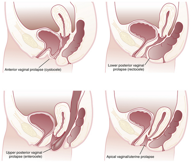
APICAL VAGINAL AND UTERINE PROLAPSE
Although vaginal prolapse can occur without uterine prolapse, the uterus cannot descend without carrying the upper or apical portion of the vagina with it.
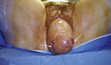
Complete procidentia (uterine prolapse through the vaginal hymen) represents failure of all the vaginal supports (Figure 23-2). Hypertrophy, elongation, congestion, and edema of the cervix may sometimes cause a large protrusion of tissue beyond the hymen, which may be mistaken for a complete procidentia. Vaginal vault prolapse or eversion of the vagina may be seen after vaginal or abdominal hysterectomy and represents failure of the supports around the upper vagina.

FIGURE 23-2 Complete uterine prolapse (procidentia). Note the lesions on either side of cervical dimple (arrows) representing pressure ulcerations from clothing/undergarments.
(Courtesy of CM Tarnay, MD, Ronald Reagan–UCLA Medical Center.)
Symptoms of POP mainly affect quality of life. However, significant sequelae of POP can occur in neglected cases of procidentia, which may be complicated by excessive purulent discharge, decubitus ulceration, bleeding, and rarely, carcinoma of the cervix.
ETIOLOGY OF PROLAPSE
The pelvic fascia, ligaments, and muscles may become attenuated from excessive stretching during pregnancy, labor, and difficult vaginal delivery, especially with forceps or vacuum assistance. Asian and black women appear less likely than white women to develop prolapse.
Increased intraabdominal pressure resulting from a chronic cough, ascites, repeated lifting of heavy weights, or habitual straining as a result of constipation may predispose to prolapse. Atrophy of the supporting tissues with aging, especially after menopause, also plays an important role in the initiation or worsening of pelvic relaxation. Iatrogenic factors include failure to adequately correct all pelvic support defects at the time of pelvic surgery, such as hysterectomy.
DIAGNOSIS
Vaginal examination is facilitated by using a single-blade speculum. While depressing the posterior vaginal wall, the patient is asked to strain down. This demonstrates the descent of the anterior vaginal wall consistent with prolapse and urethral displacement. Similarly, retraction of the anterior vaginal wall during straining will accentuate posterior vaginal defects and uncover enterocele and rectocele if present. Rectal-vaginal examination is often useful to demonstrate a rectocele and to distinguish it from an enterocele.
QUANTIFYING AND STAGING PELVIC ORGAN PROLAPSE
The preferred method to describe and document the severity of POP is the Pelvic Organ Prolapse Quantification (POP-Q) system. The extent of prolapse is evaluated and measured relative to the hymen, which is a fixed anatomic landmark. The anatomic position of the six defined points for measurement is denoted in centimeters above the hymen (negative number) or centimeters below the hymen (positive number). The plane at the level of the hymen is defined as zero (Figure 23-3).

FIGURE 23-3 Illustration showing a side view of female pelvis. Six sites (points Aa, Ba, C, D, Bp, and Ap), genital hiatus (gh), perineal body (pb), and total vaginal length (tvl) used for pelvic organ support quantitation.
(Reproduced with permission from Bump RC, et al: The standardization of terminology of female pelvic organ prolapse and pelvic floor dysfunction. Am J Obstet Gynecol 175:10, 1996.)
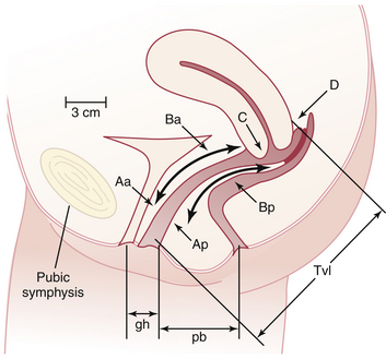
Stages of POP can be assigned according to the most severe portion of the prolapse after the full extent of the protrusion has been determined. An ordinal system is used for measurements of different points along the vaginal canal and allows for better communication among clinicians. This staging system enables more objective tracking of surgical outcomes (Table 23-1).
TABLE 23-1 PELVIC ORGAN PROLAPSE STAGING SYSTEM
Stage | Characteristics |
---|---|
0 | No prolapse |
Aa, Ba, Ap, Bp are −3 cm and C or D ≤ −(tvl −2) cm | |
1 | Most distal portion of the prolapse −1 cm (above the level of hymen) |
2 | Most distal portion of the prolapse ≥ −1 cm but ≤ +1 cm (≤1 cm above or below the hymen) |
3 | Most distal portion of the prolapse > +1 cm but > +(tvl − 2) cm (beyond the hymen; protrudes no farther than 2 cm less than the total vaginal length) |
4 | Complete eversion; most distal portion of the prolapse ≥ +(tvl − 2) cm |
Aa, point A of anterior wall; Ba, point B of anterior wall; Ap, point A of posterior wall; Bp, point B of posterior wall; −, above the hymen; +, beyond the hymen; tvl, total vaginal length.
Reproduced with permission from Harvey MA, Versi E: Urogynecology and pelvic floor dysfunction. In Ryan KJ, Berkowitz RS, Barbieri RL, Dunaif A (eds): Kistner’s Gynecology and Women’s Health, 7th ed. St. Louis, Mosby, 1999. Copyright © 1999 Elsevier.
MANAGEMENT
Prophylactic measures to mitigate the symptoms of POP include identifying and treating chronic respiratory and metabolic disorders, correction of constipation and intraabdominal disorders that may cause repetitive increases in intraabdominal pressure, and administration of estrogen to menopausal women. Failure to recognize and treat significant support defects at the time of concomitant gynecologic surgery may lead to progression of existing prolapse and the development of urinary incontinence or retention and urinary tract infections (UTIs).
Nonsurgical Treatment
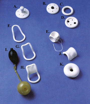
When only a mild degree of pelvic relaxation is present, pelvic floor muscle exercises may improve the tone of the pelvic floor musculature. Pessaries, which provide intravaginal support (Figure 23-4), may be used to correct prolapse by “propping up” the vagina. They can be considered when the patient is medically unfit or refuses surgery or during pregnancy and the postpartum period. They are also useful to promote healing of a decubitus ulcer before surgery for prolapse.

FIGURE 23-4 Some types of vaginal pessaries used for prolapse: Gellhorn (A), Shaatz (B), ring (C), ring with support (D), cube (E), Smith (F), Hodge (G), Hodge with support for cystocele (H), Inflatoball (I), Gehrung (J), and doughnut (K).
Pessaries require proper fitting and selection of the appropriate type and size. They should be removed, cleaned, and reinserted every 6 to 12 weeks. They may cause vaginal irritation and ulceration. Neglect may result in serious consequences, including fistula formation, impaction, bleeding, and infection.
Surgical Treatment
The main objectives of surgery are to relieve symptoms and restore normal anatomic relationships and visceral function. Preservation or restoration of satisfactory coital function when desired and a lasting operative result are also important goals.
REPAIR OF VAGINAL PROLAPSE.
Anterior colporrhaphy corrects anterior vaginal wall prolapse and helps support the urethra. It involves plication of the pubocervical fascia to support the bladder and urethra.
When the anterior prolapse involves a direct detachment of lateral vaginal support, it is considered a paravaginal defect. Paravaginal defect repairs involve exposure of the retropubic space. Interrupted permanent sutures are used to reattach bilaterally the anterosuperior vaginal sulci to the arcus tendineus fasciae (“white line”) extending from the ischial spine to the lower edge of the pubic ramus. In the presence of SUI, additional supportive measures are taken to achieve suspension of the bladder neck and proximal urethra.
Posterior colporrhaphy corrects a posterior vaginal wall prolapse and is similar in principle to anterior colporrhaphy. Site-specific posterior vaginal repairs can be performed after identification of the discrete endopelvic fascial breaks and reapproximating this thicker tissue identified during rectal examination. Perineorrhaphy repairs a deficient perineal body.
Recent modifications of these procedures involve the use of permanent suture or the addition of graft materials to augment the durability of the repair. These modifications can be accomplished using minimally invasive techniques.
REPAIR OF APICAL PROLAPSE.
When the uterus is present, hysterectomy may be performed to facilitate exposure of the apical support structures. Hysterectomy, however, is not a requirement in settings in which uterine removal is not desired. The repair of apical defects may require peritoneal entry for the repair of an enterocele. After identification of the enterocele, the contents are reduced, the neck of the peritoneal sac is ligated, and the defect is repaired by approximating the uterosacral ligaments and levator ani muscles to restore continuity in the endopelvic fascia.
Vaginal vault suspension (colpopexy) for apical prolapse is performed to secure a durable fixation point for the top of the vagina. This can be accomplished vaginally or abdominally by suspending the vaginal vault to the sacrum, sacrospinous ligaments, uterosacral ligaments, or other firm points of fixation.
VAGINAL CLOSURE PROCEDURES
For women with advanced vaginal prolapse who no longer desire coital function, there are less invasive surgical options. A LeFort colpocleisis involves suturing the partially denuded anterior and posterior vaginal walls together in such a way that the uterus remains in situ and is supported above the partially occluded vagina. In women with posthysterectomy prolapse, a complete colpocleisis involves total obliteration of the vagina. These “obliterative” procedures are traditionally reserved for elderly women who are not likely to tolerate more invasive reparative surgery.
Urinary Incontinence
Urinary incontinence is defined as the involuntary loss of urine that is objectively demonstrable and is a social or hygienic problem. Urinary incontinence has been reported to affect 15% to 50% of women. The problem increases in prevalence with age, reaching more than 50% in elderly persons in nursing homes. It is estimated that the direct financial cost of urinary incontinence in the United States is between $10 and $15 billion per year.
ANATOMY AND PHYSIOLOGY OF THE LOWER URINARY TRACT
In the adult female, the urethra is a muscular tube, 3 to 4 cm in length, lined proximally with transitional epithelium and distally with stratified squamous epithelium. It is surrounded mainly by smooth muscle. The striated muscular urethral sphincter, which surrounds the distal two thirds of the urethra, contributes about 50% of the total urethral resistance and serves as a secondary defense against incontinence. It is also responsible for the interruption of urinary flow at the end of micturition.
The two posterior pubourethral ligaments provide a strong suspensory mechanism for the urethra and serve to hold it forward and in close proximity to the pubis under conditions of stress. They extend from the lower part of the pubic bone to the urethra at the junction of its middle and distal third.
INNERVATION
The lower urinary tract is under the control of both parasympathetic and sympathetic nerves. The parasympathetic fibers originate in the sacral spinal cord segments S2 through S4. Stimulation of the pelvic parasympathetic nerves and administration of cholinergic drugs cause the detrusor muscle to contract. Anticholinergic drugs reduce the vesicle pressure and increase the bladder capacity.
The sympathetic fibers originate from thoracolumbar segments (T10 through L2) of the spinal cord. The sympathetic system has α- and β-adrenergic components. The β-fibers terminate primarily in the detrusor muscle, whereas the α-fibers terminate primarily in the urethra. α-Adrenergic stimulation contracts the bladder neck and urethra and relaxes the detrusor. β-Adrenergic stimulation relaxes the urethra and detrusor muscle. The pudendal nerve (S2 to S4) provides motor innervation to the striated urethral sphincter.
FACTORS INFLUENCING BLADDER BEHAVIOR
Sensory Innervation
Afferent impulses from the bladder, trigone, and proximal urethra pass to S2 through S4 levels of the spinal cord by means of the pelvic hypogastric nerves. The sensitivity of these nerve endings may be enhanced by acute infection, interstitial cystitis, radiation cystitis, and increased intravesical pressure. The latter may occur in the standing or bending-forward position or in association with obesity, pregnancy, or pelvic tumors.
Inhibitory impulses, probably relayed by the pudendal nerve, also pass to S2 through S4 after mechanical stimulation of the perineum and anal canal. Their passage may explain why pain in this region can cause urinary retention.
Central Nervous System
In infancy, the storage and expulsion of urine are automatic and controlled at the level of the sacral reflex arc. Later, connections to the higher centers become established, and by training and conditioning, this spinal reflex becomes socially influenced so that voiding can be voluntarily accomplished. Although organic neurologic diseases may interrupt the influence of the higher centers on the spinal reflex arc, patterns of micturition may also be profoundly altered by mental, environmental, and sociologic disturbances.
CONTINENCE CONTROL
The bladder must store and hold urine painlessly and then, at the appropriate social setting, empty urine effectively. The normal bladder holds urine because the intraurethral pressure exceeds the intravesical pressure. The pubourethral ligaments and surrounding endopelvic fascia support the urethra so that abrupt increases in intraabdominal pressure are transmitted equally to the bladder and proximal third of the urethra, thus maintaining a pressure gradient between the two structures. In addition, a reflex contraction of the levator ani compresses the mid-urethra, decreasing the likelihood of urine loss.
Stress Urinary Incontinence
SUI is involuntary leakage of urine in response to physical exertion, sneezing, or coughing.
ETIOLOGY
The most commonly accepted theory for the pathogenesis of SUI is urethral hypermobility due to vaginal wall relaxation, displacing the bladder neck and proximal urethra downward. When this occurs, increased intraabdominal pressure from coughing, sneezing, or physical exertion is no longer transmitted equally to the bladder and proximal urethra. The normal urethral resistance is overcome by this increased bladder pressure, and leakage of urine results.
The second possible mechanism is intrinsic sphincter deficiency, where the urethra fails to close in response to increases in intraabdominal pressure. This cause of SUI is analogous to having a leaky “valve” in the urethra.
Factors that contribute to SUI include childbearing, previous urogenital surgery, pelvic radiation, estrogen deficiency (menopause), and medications such as diuretics and α-adrenergic blockers.
PELVIC EXAMINATION
Inspection of the vaginal walls should be performed with a single-blade speculum, which allows optimal visualization of the anterior vaginal wall and urethrovesical junction. Scarring, tenderness, and rigidity of the urethra from previous vaginal surgeries or pelvic trauma may be indicated by a scarred anterior vaginal wall. Because the distal urethra is estrogen dependent, the patient with urogenital atrophy also has atrophic urethritis.
DIAGNOSTIC TESTS
Stress Test
The patient is examined with a full bladder in the lithotomy position. While the physician observes the urethral meatus, the patient is asked to cough. SUI is present if short spurts of urine escape simultaneously with each cough. A delayed leakage, or loss of large volumes of urine, suggests uninhibited bladder contractions. If loss of urine is not demonstrated in the lithotomy position, the test should be repeated with the patient in a standing position.

Full access? Get Clinical Tree

