Chapter 62 Genetics
Our understanding of human genes and of the genetic basis of disease has grown dramatically, especially with the completion of mapping of the Human Genome Project. Molecular technology for identifying human genes has allowed for the diagnosis and understanding of the mechanisms of human diseases. Medical genetics involves not only the diagnosis, treatment, and management of individuals with multiple congenital anomalies, developmental disabilities, and hereditary diseases, but it also involves human disorders having complex inheritance, such as diabetes, many types of cancer, and heart disease (e.g., cardiomyopathy). Knowledge of medical genetics is relevant to all medical practice, particularly pediatrics.
What Are the Reasons for a Genetic Consultation?
Children and their families are most commonly referred for clinical genetics evaluation because of birth defects or a known or suspected genetic disorder. Examples include the following:
Growth problems, including short stature, failure to thrive, or overgrowth
Neurodevelopmental problems, such as mental retardation, developmental regression, seizures, ataxia, or behavioral problems
Premature or delayed puberty or ambiguous genitalia
Prenatal exposure to alcohol, drugs, or other teratogens
Recurrent miscarriages or advanced maternal age
Family history of a known disorder, such as cystic fibrosis or fragile X syndrome
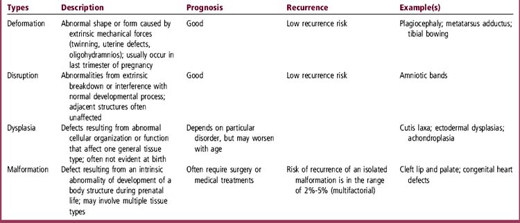
BIRTH DEFECTS
ETIOLOGY
How Common Are Birth Defects?
A birth defect that interferes with normal function of the affected tissue or organ is identified in approximately 3% of all children within the first year of life and in 5% by adolescence. Several studies have indicated that congenital abnormalities account for about 17% to 27% of all hospitalizations for pediatric patients.
What Causes Birth Defects?
Birth defects and genetic disorders may be caused by (1) single-gene mutations, (2) chromosome abnormalities, (3) multifactorial factors, the interaction between genes and environment, and (4) teratogens.
Single-gene disordersmay have autosomal dominant, autosomal recessive, X-linked recessive, or X-linked dominant inheritance. There are now more than 5000 such disorders recognized. It is estimated that single-gene disorders affect 2% of the general population over a lifetime and 6% to 8% of hospitalized children.
Chromosome disordersaffect almost 1% of live-born children and account for approximately 50% of spontaneous abortions. These involve both numerical and structural chromosome defects.
Multifactorial inheritancemay account for up to 60% of disease in the entire population in a lifetime. Common disorders in adults and isolated congenital anomalies in children result from the combined effects of genetic mutation and environmental factors.
Teratogenscause morphologic and/or functional abnormalities in a fetus. They include infectious agents, physical agents, drug and chemical agents, and maternal metabolic and genetic factors. The effects of teratogens are preventable in the absence of the offending factor.
EVALUATION
How Do I Evaluate a Child with Multiple Birth Defects?
The history for a child with multiple congenital anomalies should specifically emphasize prenatal events. These include (1) maternal factors such as diabetes, hypertension, exposures to prescription and other drugs, uterine abnormalities, and others and (2) fetal factors including abnormal fetal movements, ultrasound findings in the fetus, and amniotic fluid analyses. Ask about development, behavior, growth patterns, and presence of impaired hearing or vision. The family history should include information on ethnic origin, consanguinity, similar abnormalities in other family members, and recurrent miscarriages or stillbirths. Parental age is also important because advanced maternal age is associated with risk of chromosomal abnormalities in the fetus, and advanced paternal age is associated with risk of new autosomal dominant mutations.
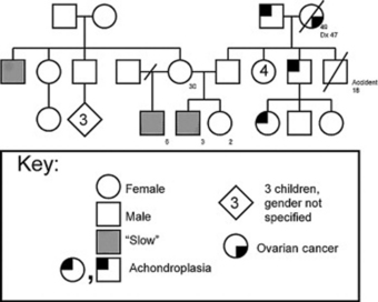
How Do I Develop a Family Pedigree?
A family pedigree is essential to help determine whether an identifiable mode of inheritance can explain the presenting problem: autosomal dominant, autosomal recessive, X-linked, or chromosome translocation. The family history is recorded in a pedigree with symbols representing individuals and their symptoms or disorders. At least three generationsare needed to establish inheritance patterns. If the pedigree shows male-to-male transmission, an equal number of males and females affected, and affected individuals in each successive generation, this confirms autosomal dominant inheritance.If males are predominantly affected on the maternal side of the family and vertical transmission is demonstrated, then X-linked inheritanceis likely. Horizontal patterns of affected individuals or siblings, or consanguinity (relatedness), suggest autosomal recessive inheritance.The family history is recorded in a pedigree with symbols representing individuals and their symptoms or disorders (Figure 62-1).
How Do I Recognize a Birth Defect?
An anomaly can be either a minor or a major structural abnormality. Minor anomalieshave no serious medical or cosmetic effects. They serve as clues to the diagnosis of a specific pattern of malformation. Examples of minor anomalies include preauricular ear pits, fifth finger clinodactyly, single palmar crease, and epicanthal folds. The presence of two or more minor malformations should “raise a red flag” and warrants a search for an underlying major malformation. Major malformationsusually require surgical or medical intervention and can reduce life expectancy if not corrected. Examples of major anomalies include microphthalmia, hydrocephalus, gastroschisis, and clubfeet.
What Is Important on Physical Examination?
The physical examination must look for minor and major structural anomalies and specifically note the physical growth of various parts of the body such as upper and lower segment measurements or size of the ears. This requires careful measurements to provide quantitative assessment and comparison with established norms. There are published graphs of these measures (Hall et al., 1995).
Is Testing Needed for an Isolated Anomaly?
An isolated birth defect or major anomaly is most likely caused by multifactorial inheritance but could be familial with autosomal dominant inheritance. In most cases, further testing cannotconfirm this mode of inheritance. Parents should be counseled that the risk of recurrence of the anomaly in future offspring is approximately 2% to 5%. In some cases, gene mutations may have been identified as the cause of a particular malformation. An example is coronal craniosynostosis, in which mutations in the fibroblast growth factor receptor 3 (FGFR3)gene may be causative. Because many of the major malformations have associated minor anomalies, it may be warranted to obtain routine chromosome analysis when evaluating such a child.
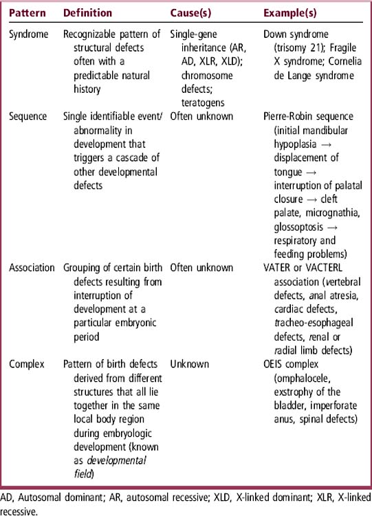
Do Multiple Birth Defects Have Recognizable Patterns?
Multiple major and minor structural anomalies can have recognizable patterns: syndrome, sequence, association, and complex. Table 62-2defines and gives examples of each pattern. Each of these patterns has implications for etiology, natural history, and prognosis. The patterns of malformationmust be considered when a child has multiple major and minor anomalies, especially if accompanied by growth, developmental, or behavioral abnormalities. Each pattern may have different modes of inheritance: single gene, chromosome abnormality, or multifactorial causes.
What Is the Evaluation for Multiple Birth Defects?
The child with two or more minor anomalies should undergo an evaluation for underlying major anomalies, because many are occult. A meticulous physical examination and selected imaging studies and laboratory tests can help identify associated birth defects or medical problems. For instance, a child who has preauricular pits or protruding ears (minor anomalies) should undergo hearing testing and renal ultrasound studies to search for the hearing loss and renal malformations found in the branchio-oto-renal (BOR) syndrome. Complications associated with suspected disorders should also be sought using imaging and/or laboratory studies. For example, a child suspected of having Williams syndrome (caused by microdeletion of chromosome 7q11) is at risk for hypercalcemia, renal artery stenoses and/or calculi, supravalvular aortic stenosis, and pulmonic stenosis. Surveillance testing with total and ionized serum calcium levels, electrolytes, blood urea nitrogen (BUN) and creatinine, urinalysis, renal ultrasound, and echocardiogram may identify abnormalities. Other birth defects may justify chromosome analysis, fluorescence in situ hybridization (FISH) analysis, and DNA testing.
When Should Chromosome Analysis Be Done?
Chromosome analysis is recommended for the child with multiple congenital anomalies, developmental delay, and/or growth abnormalities, as well as for couples with multiple pregnancy losses. A normal karyotype or chromosome analysis consists of 23 pairs of chromosomes, including 22 pairs of autosomes and 1 pair of sex chromosomes (46,XX in females, and 46,XY in males). Microdeletions and microduplications are detected by chromosomal microarray analysis, which targets telomeres, centromeres, and known microdeletion/microduplication syndromes.
What Is the Fluorescence In Situ Hybridization Test?
Very small deletions or duplications detected by chromosome microarray analysis are confirmed by FISH. This study is useful to identify conditions caused by “microdeletions,” such as DiGeorge/velocardiofacial syndromes (chromosome 22q11 deletion) and Williams syndrome (chromosome 7q11.2 deletion).
What Must I Do before Requesting DNA or Molecular Tests?
You must make a precise and correct clinical diagnosis for a patient with multiple birth defects, mental retardation, or metabolic abnormality before considering DNA or molecular testing. These tests can be used only if there is a known gene mutation. In addition, there must be a clinical and/or research laboratory available to perform the tests. Most genetic disorders do not have an identified gene mutation, so DNA testing is not available.
What Is DNA Testing?
DNA testing detects gene mutations or changes in the DNA sequence as small as a single nucleotide or as large as thousands of base pairs. These are too small to be detected by chromosome or FISH analysis. Molecular techniques can detect gene mutations for many different genetic disorders.

Stay updated, free articles. Join our Telegram channel

Full access? Get Clinical Tree

