40 Genetic Disorders
The science of the human genome is transforming the field of genetics as we understood it from a mendelian inheritance perspective into a new genomics paradigm. In the new paradigm the focus is no longer on single genes and chromosomes, but on the influence of individual genes, the effects of many genes working in concert with one another, and with environmental exposures via epigenetics pathways to influence health outcomes. The issues involve identification of genome sequences of interest, interpretation of how those genes affect health, and use of that information to improve the health of children and their families (McBride and Guttmacher, 2009).1
Genetic factors are linked to many disorders found in children. Some of these disorders are considered to be classic genetic diseases. Examples include cystic fibrosis, Down syndrome, and Duchenne muscular dystrophy. However, many of the most prevalent disorders of childhood have some genetic components. The chance that a child has or will develop a single-gene, chromosomal, or malformation condition during his or her life is between 3.2% and 7.3% (Jorde et al, 2006). The Human Genome Project is helping researchers identify the genetic markers for many more conditions. With this knowledge the intersection between genetics and environment (i.e., the field of epigenetics) is becoming clearer, and newborn screening has expanded, allowing for earlier identification of genetic conditions, and new therapies are being developed.
The manifestations of genetic diseases can appear immediately after birth or after many years, such as in patients with Huntington chorea; and can present in biochemical, reproductive, growth, developmental, or behavioral ways. Therefore, the primary health care provider must be constantly vigilant for the possibility of genetic disease. Furthermore, once a genetic condition is suspected, referral to a medical geneticist is not always required or possible. Primary care providers need to be knowledgeable, yet know their own limitations and set personal criteria for referral to specialists.
Caring for children with significant long-term problems confers enormous responsibilities on the provider, family, community, and society. Primary care providers assume a variety of roles related to the care of children with genetic disorders. These roles include promoting health of individuals with genetic conditions, assisting children and families to reduce risk for medical problems by helping families make decisions about childbearing, screening for early detection to prevent disability, assisting parents to use specialized services, teaching health principles, monitoring and evaluating clients with genetic diseases, and working with families under the stress of caregiving. Ethical decision-making has a particularly important role in the area of genetics and genetic counseling.
Cellular and Molecular Genetics
Humans have 46 chromosomes arranged in 23 pairs. Twenty-two pairs are autosomes (the same in males and females) and are homologous because their deoxyribonucleic acid (DNA) is very similar. The remaining pair is the sex chromosomes, with two X chromosomes for females and one X and one Y chromosome for males. They are not homologous. Each chromosome has a long arm (q) and a short arm (p) and is numbered according to its distinct appearance from the largest to the smallest. The gametes (egg and sperm) have half the chromosome complement from the parents (23). During meiosis (formation of the haploid with 23 chromosomes from the egg or sperm), the original paired chromosomes from paternal and maternal sides cross over and exchange genetic material, resulting in genetic diversity. Fusion at fertilization restores the 46-chromosome (23-pair) complement, with one of each chromosome pair from each gamete. As a result of genetic variation, a gene may differ from one individual to another in its DNA sequence. These differences in sequencing are called alleles. If the two alleles at any given location of a pair of homologous chromosomes are identical, the locus is homozygous. If the two alleles are different, the locus is heterozygous.
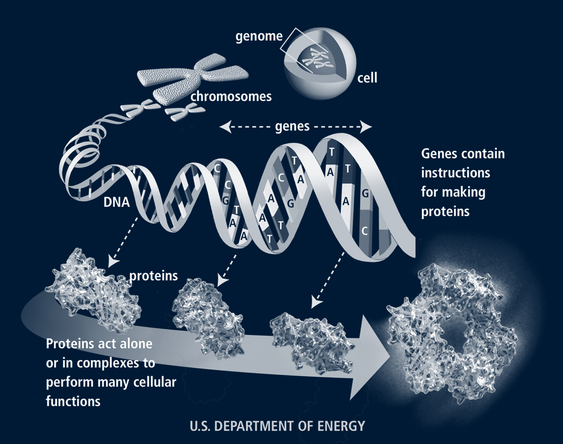
Genes, which carry the information about inherited characteristics from parent to child, are arranged linearly on the chromosomes, each with a specific locus. Thousands of genes are located on each chromosome. Not all genes are active at once; certain mechanisms activate them at various developmental points. In homozygous loci, the genes from a pair of chromosomes carry similar instructions regarding the trait of interest; in heterozygous loci, the instructions are different for each gene. In the latter case, one gene may be dominant, with its instructions manifested in the phenotype, as in Huntington disease, or the genes can be co-dominant, as in the case of individuals with blood type AB.
The human genome contains approximately 25,000 to 30,000 genes. Genes are composed of DNA. Each DNA molecule includes pairs of nitrogenous bases—adenine, cytosine, guanine, and thymine (labeled A, C, G, T)—wound around a histone protein core in a double helix. There are more than 3 billion base pairs in the human genome for an individual. From the four nitrogenous bases, 64 triple-base combination sequences (codons) of A, C, G, and U (uracil is substituted for thymine in the messenger ribonucleic acid [mRNA] at this point) such as GUA, UUG, and CGG are possible (Jorde et al, 2006). Three codons signal the end of a gene (stop codons) and 61 define the 20 amino acids. Thus each amino acid may be specified by more than one codon. The sequence of the amino acids directs the synthesis of proteins in the cell cytoplasm (Fig. 40-1). Telomeres at the tips of each chromosome protect the chromosome from breaking down. These deteriorate with age.
Now that the genome has been defined, geneticists are working to understand the functions of individual genes. In the case of conditions such as Down syndrome, which has been identified as a chromosomal disorder, understanding of the functions of the many genes on chromosome 21 may help clarify how some of the various phenotypes of the condition are controlled and, ultimately, might be able to provide therapies for some of the problems such as leukemia or cataracts, which emerge later in life (Patterson, 2009). Similar progress may offer new perspectives and options for other genetic conditions.
Epigenetics
Epigenetics is defined as the study of heritable changes in genome function that occur without a change in DNA sequence. This includes the study of how patterns of gene expression are passed from one cell to its descendants, how gene expression changes during the differentiation of one cell type into another, and how environmental factors can change the way genes are expressed (Bagot and Meaney, 2010; Epigenome Network of Excellence, 2009).
In the nucleus of nondividing cells, genomic DNA is highly folded and compacted with histone and nonhistone proteins into a polymer called chromatin (DNA does not normally look like the nice double helix strands seen in many diagrams). The epigenome is the group of proteins around the genome that tells genes when to turn on and off. An analogy could be made to computers: the genome is the computer’s hardware; the epigenome is the software that tells it what to do.
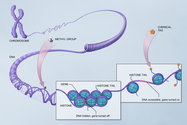
There are several recognized mechanisms for epigenetic inheritance, not all of which are discussed here. DNA methylation is the most studied. When DNA material is methylated, the gene is turned off. Methylation is involved with cellular differentiation in utero but the same process occurs throughout life. A second mechanism for epigenetic modifications to DNA functioning involves the way DNA attaches to histones. It is called chromatin modification. DNA is wrapped around a series of proteins called histones. If these proteins are tightly joined to the DNA, the DNA is hidden from exposure and cannot express itself. If the histone wrap is loosened, often through acetylation, enzymes, or certain forms of RNA, then the gene may be expressed (Fig. 40-2). In general, when chromatin is tightly folded, gene expression is restricted while more open chromatin allows gene expression. In other words, gene regulation varies depending on the type of histone linkage. Certain RNA proteins are also transmitted with reproduction and are involved with epigenetic inheritance (Jablonka and Raz, 2009).

FIGURE 40-2 Epigenetic diagram. Chromatin modification: deoxyribonucleic acid (DNA) is wrapped around a series of proteins called histones. If these proteins are tightly joined to the DNA, the DNA is hidden from exposure and cannot express itself. If the histone wrap is loosened, often through acetylation, enzymes, or certain forms of ribonucleic acid (RNA), then the gene may be expressed. Methyl groups attached to the DNA also affect gene expression.
(From The National Institutes of Health Common Fund, Division of Program Coordination, Planning, and Strategic Initiatives, National Institutes of Health.)
Genes are regulated when the histone allows signals to reach the gene. These signals may come from one cell touching another as in neurologic growth, when a cell releases factors that are picked up by neighboring cells as happens at cell synapses, through hormones that are broadcast to the whole body (cells that are tuned in will respond), or by signals coming from environmental factors. Some of these environmental factors reach cells directly and others indirectly (mediated, for example, by stress), and cause responses that are transmitted across body systems. Signals are passed to a gene regulatory protein that attaches to a specific sequence of DNA molecules. Once the protein is attached to the DNA molecule, the gene turns off. The epigenetic tags allow the cell to “remember” what to do over time and over many replications. Some tags are passed on to later generations. Certain enzymes can be recruited to remove epigenetic tags, the histones, or both, and cells are stripped of many of their tags with reproduction of sperm and egg. The tags that remain to be passed along to the next generation are referred to as imprinted. More than 80 genes can be imprinted (Weinhold, 2006). Chemical exposures can cause methylation of DNA and transmission to future generations—altering the function of cells but not the DNA itself (Jablonka and Raz, 2009).
An individual normally has one copy of an imprinted gene (it came along on the sperm or egg chromosome). Improper imprinting can cause an individual to have two copies of active, imprinted genes, or two inactive copies. Prader-Willi and Angelman syndromes come from the same imprinting area on chromosome 15. If the individual is missing gene activity that normally comes from the father or there are two active copies from the mother, Prader-Willi will result; if the imprinting defect results in failed gene activity that normally comes from the mother, Angelman will result.
The study of epigenetics is revolutionizing our understanding of many conditions (e.g., autism spectrum disorders, which are known to have both genetic and environmental etiologic factors; epigenetics may explain how the various factors interrelate to cause these disorders). Rett, fragile X, Prader-Willi, Angelman, and all demonstrate dysregulation of normal epigenetic mechanisms (Grafodatskaya et al, 2010). It is thought that many cancers may result from epigenetic control of gene expression rather than defective genes themselves, or may be caused by a mixture of the two factors. Cancer epigenetics may involve disruption of the stem cells, sometimes when they have replicated many times over years and no longer seem to be able to function properly (Feinberg, 2007; NOVA, 2007). Some of the endocrine disruptor toxins in the environment cause epigenetic trangenerational effects, meaning that the germline is affected and this change is transmitted to future generations (Guerrero-Bosagna and Skinner, 2009).
Epigenetic research has contributed to the development of new epigenetic therapy in which instructions to cells are changed, allowing them to reactivate in a normal way after having been silenced by cancer cells (Issa, 2010).
Ethical Issues
Since 1990, largely due to the work of the Human Genome Project (HGP), the technical capability to diagnose a hereditary condition for those who are presymptomatic or currently symptomatic, to identify those who are carriers of a genetic condition, and to determine susceptibility to a genetic condition has increased dramatically. However, the availability of this technology raises significant ethical issues. The HGP was concerned about these issues from the beginning and has a branch specifically devoted to oversight of these concerns (the Ethical, Legal, and Social Initiative [ELSI]). Issues involve the rights to privacy and confidentiality, rights to know and right not to know, and whether there is a duty to warn third parties (Ross, 2008).
Maintenance of confidentiality is a challenge. The presence of genetic information in the medical record, health insurance diagnostic database, or in DNA databases such as newborn screening specimens mandates policies to maintain the confidentiality and integrity of these records. The Health Insurance Portability and Accountability Act (HIPAA) of 1996 specifies the duty for clinicians not to disclose medical information without the signed consent of the patient or the child’s parents (U.S. Department of Health and Human Services [USDHHS], 2010).
As genetics tests are conducted, it is also possible to discover unanticipated information, for instance, data regarding the parentage of the child being tested. Situations such as misattribution of paternity and children being raised by nonbiologic parents may be discovered. There is disagreement about whether these findings should be routinely disclosed. Prior agreement in the consent process can assist in the decision about whether to disclose this information.
The ability to screen for a large number of genetic disorders via the tandem mass spectrometry newborn screening process has increased the number and types of conditions that can be included. Because these programs are organized by and paid for through state governments, there is some variability in required tests between states. Congenital hypothyroidism, sickle cell disease, sickle-C, sickle-beta thalassemia, classical galactosemia, and phenylketonuria are mandated in newborn screening programs in all U.S. states. An additional sixty-one conditions are included in individual state newborn screening programs (National Newborn Screening & Genetics Resource Center, 2006). Three principals have been suggested by the Institute of Medicine (IOM) to be used in making decisions about the introduction or continuation of tests:
• Identification of the genetic condition must provide a clear benefit to the child.
• A system must be in place to confirm the diagnosis.
• Treatment and follow-up must be available for affected infants.
Predictive genetic testing is another area for ethical consideration. The availability of presymptomatic testing for conditions that may not become apparent until adulthood, such as Huntington disease and breast cancer, has raised another set of questions. Should children and adolescents be tested for such conditions? Arguments related to the issue of predictive genetic testing generally fall into four categories: potential provision of good news (i.e., the child is found not to have the genetic markers); unbearability of knowing (if the child has the markers); identity and adjustment (if the child has the markers); and parental anxiety and uncertainty (Borry et al, 2008). Generally it is recommended that genetic testing for late-onset conditions be deferred until adulthood when individuals, rather than their parents, can make the decision, unless there is evidence that early diagnosis can result in treatment strategies that will alter the progression of the disease (American Academy of Pediatrics [AAP] Committee on Bioethics, 2001). If predictive testing is considered, parents should have the right to decide whether to have their child tested and to receive counseling regarding the disease including diagnostic workup, ongoing research, associations, resources available, and clinical trials in progress or registering for trials in the future (Trott and Matalon, 2009).
Biobanks including many thousands of genetic samples from children are being collected to study genetic-environmental influences on disease. Should participants have access to the data? To what extent do those children have a right to privacy?
Families also face issues of disclosure of genetic information within their own families. What information should be disclosed? When? To whom? Disclosure is influenced by the perceived risks and benefits of doing so, the sense of closeness among the family members, concerns about reactions from those receiving the information, their sense of personal risk, and readiness to disclose information (Gallo et al, 2009). Disclosure to children is also an issue many families face. Families worry about psychological harm, the child’s lack of autonomy in deciding whether to be tested, and concerns that the child won’t understand the information given. The child’s developmental age is a key factor. For preschoolers there may only be a dawning awareness of symptoms, treatments, or physical differences. Parents will want to choose whether they disclose information, choose to wait, or want a health care professional to begin the dialogue with the child about the genetic condition of interest. School-age children may question parents about the implications of a particular diagnosis or become aware of reproductive risks. Adolescents are aware of conditions and want information in more depth. They may have very specific questions about reproductive risks. Health care professionals need to assist patients and families to disclose information as is appropriate, Questions such as, “Have you ever talked with your child about his condition?” may start a helpful discussion.
These are only a few of the ethical issues under discussion. Primary care providers must be part of the ongoing debate and be aware of the issues, policies, and laws as they work with families who are trying to make decisions.
Genetics Testing in the Future
Genetics testing will become more complex in the future and primary care providers will need to be knowledgeable in all these areas. First, universal screening can be expected to expand as therapies evolve, making it possible to prevent or modify significant pathology in affected individuals. Second, screening will be expanded for diagnosis of common conditions. For example, more than 50% of sensorineural hearing loss is due to genetic conditions. When infants are identified through newborn hearing screening, they will then undergo genetic testing to ascertain possible causes. Some autism and obesity cases are now connected to certain genetic profiles in children. Hypertension, diabetes, heart disease, and cancer are adult conditions in which greater genetic information will affect identification of and management of those at risk (Lose, 2008). Third, additional screening will be done to identify those at risk for future problems, such as the breast cancer and Parkinson disease tests that are already available. Finally, pharmacogenetics will involve genetic testing to ascertain drug responses in individuals with different genetic profiles (Cheng et al, 2008). Primary care providers will serve as translators for patients needing and receiving genetic information over their lifetime (Lose, 2008).
Causes of Genetic Variation and Genetic Disorders
Mutations occur when genetic material is permanently changed through alteration, deletion, duplication, or misplacement. Sometimes mutations arise spontaneously, but once the change occurs in the germ cells, it is transmitted to future generations. Mutations are defined as characteristics being present in less than 1% of the population. A change in greater than 1% is called a polymorphism. Mutations and polymorphisms may be benign, beneficial, or detrimental.
Genetic disorders are classified as chromosomal disorders, in which the entire chromosome or large segments of it are duplicated or missing; single-gene disorders, in which single genes are altered; and multifactorial problems, in which multiple genetic and environmental factors interact. Although the majority of genetic conditions fit in these categories, other patterns of inheritance, such as mitochondrial inheritance may lead to genetic disorders. Conditions that result from mutation in mitochondrial DNA are inherited through the maternal line. Some epigenetic changes can also be inherited, at least to the next generation and perhaps more. This is an emerging area of research.
Chromosomal Disorders
Chromosomal disorders are present in 0.6% to 0.9% of the general population (Jorde et al, 2006). There is also a high frequency of chromosomal disorders in spontaneous abortions and stillbirths. Such disorders are commonly linked to alterations in cognitive development; linear growth, usually short stature; and congenital anomalies. The chromosomal disorders include problems of chromosome number (increase or decrease in the number of chromosomes), structure, or both.
The prevalence of chromosomal disorders due to nondisjunction (failure of homologous pairs to separate properly during meiosis) increases with advancing maternal age. Testing to diagnose a chromosome disorder is done through cell culture and chromosome analysis. In addition to the more traditional method of karyotype analysis, in which the total number of each chromosome is identified, staining techniques to identify chromosomal banding assist in the identification of deletions and duplications of chromosomal material. Techniques such as fluorescence in situ hybridization (FISH) provide the ability to identify missing, additional, or rearranged chromosomal material for some of the more common abnormalities but must be ordered for the specific location of interest.
Single-Gene Disorders
Mendelian theory describes four patterns of inheritance: autosomal dominant, autosomal recessive, X-linked dominant, and X-linked recessive. Dominant inheritance disorders occur in heterozygotes, where one gene dominates its counterpart from the other parent. Recessive inheritance disorders occur only when a person is homozygous, when the gene with a disease-causing mutation appears on both of the chromosomes of the pair. However, genetic mutations for some conditions have reduced penetrance, in which a person may have the affected gene (genotype) without expressing the observable characteristics (phenotype), and variable expressivity, in which the severity of the disease condition varies greatly. The result is that some children have clinically severe disease, whereas others, with mutations in the same gene, are more mildly affected.
Multifactorial or Multiple-Gene Disorders
Multifactorial problems result from the complex interaction of multiple genes in various sites and/or interaction of genes with the environment. Several terms are used for this group of conditions (e.g., multifactorial, multiple gene, and polygenic). Cleft lip and palate, spina bifida, hypertension, schizophrenia, pyloric stenosis, diabetes, hypercholesterolemia, Hirschsprung disease, and asthma fall into this category.
Multifactorial problems are more likely to cluster in families. The exact recurrence risk is more difficult to predict because the precise genetic and environmental risks are usually not known. However, in general, the risk for the condition increases if more family members are affected, and if the disease has a more severe expression. It is likely that epigenetics will provide explanations for many of the multifactorial disorders, identifying those combinations of genes, gene mutations, or expression or silencing of genes that result in disease.
Nontraditional Inheritance
Three additional patterns of transmission of genetic material from generation to generation have been identified—germline mosaicism, uniparental disomy, and mitochondrial inheritance.
Germline Mosaicism
In this pattern a mutation occurs in a cell of the developing organism sometime after fertilization. As cells multiply, some begin to reproduce with the mutation, whereas others do not. The outcome is a person with “mosaicism”—some normal and some abnormal cells. Whether the gametes are affected will dictate inheritance to the next generation, and the cells involved determine whether the condition is clinically relevant. Thus the term germline mosaicism is used to indicate inheritability of the trait. With germline mosaicism, parents appear normal but have some gametes with the gene mutation. The challenge, clinically, is to identify the condition as inheritable. If normal-appearing parents have a first child with a condition such as achondroplasia, which is normally autosomal dominant, the clinician would deduce that the achondroplasia was not inherited in an autosomal dominant manner (in which case one parent would have had the disorder), so a new mutation, and the possibility of germline mosaicism, must be considered. If germline mutation is present, the risk of recurrence in a second offspring is increased. It is because of such situations that genetic counseling for parents is important to help determine the risk to subsequent children.
Uniparental Disomy
Generally, children receive one chromosome from each parental pair at the time of fertilization. If, by some chance, the child receives two copies of one chromosome of a pair from one parent and none from the other parent, uniparental disomy has occurred. The result is that the child will be homozygous for every gene located on that chromosome, which increases the possibilities of an autosomal recessive disorder in the child. The process has been described in some patients with cystic fibrosis. The same process also may result in either Prader-Willi or Angelman syndrome, diseases that involve the same gene loci but differ depending on whether the copies are from the mother or the father. Beckwith-Wiedemann and Russell-Silver syndromes are also examples of uniparental disomy.
Mitochondrial DNA Inheritance
Mitochondria in cells also have DNA (mtDNA). Unlike chromosomal DNA, mtDNA is circular. All inherited mtDNA comes from the ovum—thus it has a maternal transmission pattern. Because each cell has more than one mitochondrion, there are more opportunities for mutations and also for variable expressivity; if many normal mitochondria are present, the effects from the aberrant mtDNA may be minimal. Several biopsies of different tissues will be subjected to both enzymatic and DNA analyses for diagnosis of mtDNA-related diseases. Although rare, mitochondrial diseases do play a role in some more common conditions such as deafness and non–insulin-dependent diabetes (Jorde et al, 2006).
Teratogens
Although not strictly genetic in origin, teratogens are often discussed with genetic disorders because the differential diagnosis includes factors that affect the embryo after fertilization and those that affect the DNA of the germ cells or their joining with fertilization. Fetal alcohol syndrome is an example of a condition in this category. Viral diseases, such as rubella, certain drugs, and environmental toxins—such as mercury—are also considered teratogens. The Pregnancy Exposure InfoLine (www.thepeil.org) contains current information on potential teratogenic effects of specific environmental substances.
Assessment
Primary care providers identify possible genetic disorders by using the same skills as those for other pediatric health problems: knowledge of risk factors, collection of a good history, and a complete physical examination augmented with appropriate laboratory or other studies. After the assessment, providers determine the operative genetic mechanism and develop and implement a plan of care for the patient and family with consideration of individual, family, and cultural factors. Box 40-1 identifies some common features of children or family members with genetic disorders that should lead providers to explore issues of possible genetic problems.
Risk Factors
Risk factors that may be identified in the child or family members include the following:
• Family history of known genetic disorder or recurrent pathologic condition
• Delayed development of secondary sex characteristics
• Affective disorders (e.g., schizophrenia)
• Developmental delays or learning problems
• Repeated spontaneous abortions or stillbirths
• Maternal factors, including alcohol or drug exposure, medication exposure, age older than 35 years, environmental or occupational toxin exposure
TABLE 40-1 Genetic Risks Associated With Ethnic Background
Ethnic Background | Genetic Disorder at Higher Risk |
---|---|
Northern European | Cystic fibrosis, phenylketonuria |
Jewish (Ashkenazi descent) | Tay-Sachs, Canavan, Gaucher |
West African | Sickle cell, sickle cell–hemoglobin C |
Mediterranean | Beta-thalassemia, sickle cell |
French-Canadian | Tay-Sachs, branched-chain ketoaciduria |
History
The history of genetic diseases usually includes the following main areas (Dolan and Moore, 2007):
• Family history of the disease using a pedigree format. A family history is needed to identify family members with conditions that may be genetically transmitted. The pedigree provides a visual map of the occurrence of specific traits and identifies other family members who might be at risk. Providers can see the potential pattern of inheritance and the relationships among affected family members. Past and current health of each person in the pedigree, birth histories of other family members, and mental retardation or learning problems of family members are all important areas to explore. Consanguinity should be noted; persons who have a common ancestor may each be carriers of a gene mutation present in that family.
• Environmental and occupational history. The environmental and occupational history may provide information about specific teratogenic factors that might be involved.
• Reproductive history. The mother’s reproductive history may give information about malformations, genetic conditions, or infectious diseases transmitted to other offspring. Her pregnancy and delivery of the child in question may give other information to determine whether the condition was the result of genetic factors or whether the condition was a result of trauma, infection, or some other factor occurring during the pregnancy or delivery.
• Medical history of the child
Figures 40-3 and 40-4 illustrate the pedigree notation format. Screening questions for genetic disorders that should be asked of all patients are included in Table 40-2. Families can be encouraged to record their own family history information by using programs such as the U.S. Surgeon General’s Family History Initiative (USDHHS, 2005). Parents and children should be encouraged to learn about the health of members of their family, which may, in turn, provide clues as to specific health risks for themselves. When a genetic disorder is suspected, the history must become more specific, as outlined in Table 40-3.

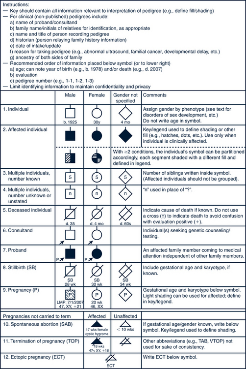
FIGURE 40-3 Pedigree model. Common pedigree symbols, definitions, and abbreviations.
(Adapted from Bennett R, French KS, Resta RG: Standardized human pedigree nomenclature: update and assessment of the recommendations of the National Society of Genetic Counselors, J Genet Counsel 17:424-433, 2008.)

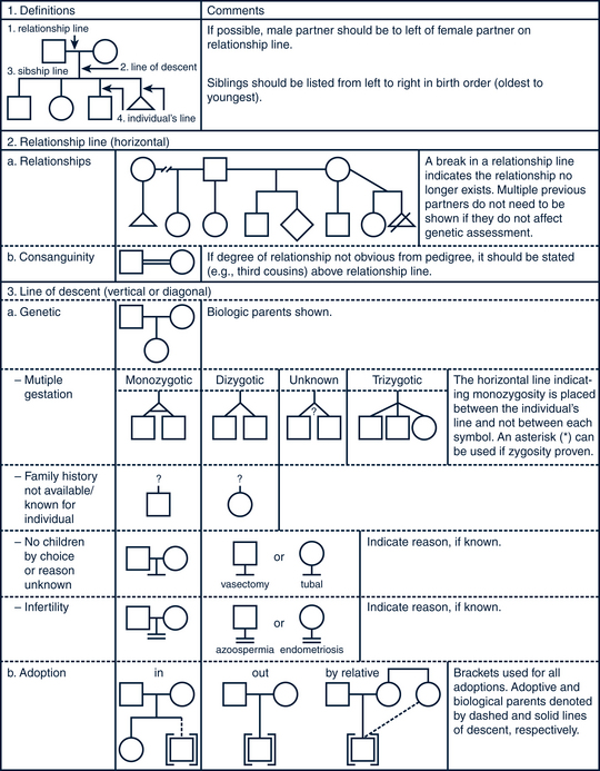
FIGURE 40-4 Pedigree line definitions.
(Adapted from Bennett R, French KS, Resta RG: Standardized human pedigree nomenclature: update and assessment of the recommendations of the National Society of Genetic Counselors, J Genet Counsel 17:424-433, 2008.)
TABLE 40-2 General Screening for Genetic Conditions: The History
Question | Rationale/Comments |
---|---|
Has anyone in the family had a birth defect? | To identify conditions that affect others in the family. If answer is yes, try to get more information about the nature of the defect. |
Is there anyone in the family with a stillborn baby or baby who died early in life? | To identify unrecognized genetic disorders. Babies who died very early may have inheritable metabolic disorders.Distinguish from sudden infant death syndrome. |
Is there any chance that you and your partner are blood-related? Is this pregnancy a product of incest? | Consanguinity of partners closer than first cousins is a risk factor for autosomal recessive disorders. If yes, recommend genetics consultation. |
Are there any diseases or traits that run in your family? | Significant if early onset, two or more close relatives affected.Genetic heart disease and genetic cancer risks are important. If yes, recommend genetic consultation and monitoring. |
Have you or any of your parents or siblings had three or more miscarriages? | May indicate a chromosome translocation. If yes, order a karyotype of the mother or father (or both). |
Does anyone in the family have mental retardation? | Look for multiple members affected and associated with dysmorphic features. If yes, recommend genetic consultation. |
What is your ethnic background? Your partner’s? | Consider ethnic risk factors and screen if at risk. |
TABLE 40-3 Specific Genetic History Questions