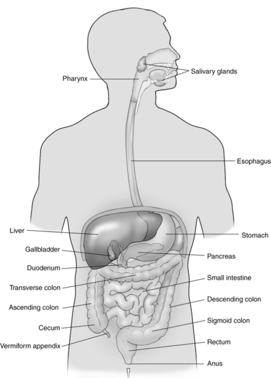
CHAPTER 14 opening at the upper end of the stomach. semiliquid mass composed of food and gastric juices. inflammation of the intestines; occurs most frequently in the ileum. a hard compacted mass of feces in the colon. failure of the stomach to empty; caused by a decrease in gastric motility. greater curvature of the stomach longer, convex, left border of the stomach. a recess or sacculation demonstrated in the walls of the ascending and transverse colon. obstruction of the small intestines. prolapse of one segment of bowel into the lumen of an adjacent segment of bowel. lesser curvature of the stomach shorter, concave, right border of the stomach. an anomalous sac protruding from the ileum; caused by an incomplete closure of the yolk stalk. situated midway between the umbilicus and the right iliac crest. extreme pain or tenderness over McBurney point; associated with appendicitis. distention of the appendix or colon with mucus. a protein-digesting enzyme produced by the stomach. opening at the lower end of the stomach. spasm of the pyloric sphincter; associated with pyloric stenosis. ridges or folds in the stomach lining. abnormal twisting of a portion of the intestines or bowel, which can impair blood flow. • Muscular tube extending from the pharynx to the stomach. • Courses down the chest through the esophageal hiatus of the diaphragm, terminating at the cardiac orifice of the stomach. • Wall layers from the outer layer to the lumen include: • Extends from the terminal ileum to the anus. • Secretes large quantities of mucus. • Divisions include the cecum, appendix, ascending colon, transverse colon, descending colon, sigmoid, rectum, and anus. • Bacteria in the colon produce vitamin K and some B-complex vitamins. • Wall layers from the outer layer to the lumen include: • Located to the left of midline, posterior to the left lobe of the liver and anterior to the abdominal aorta. • Right margin is contiguous with the lesser curvature of the stomach. • Left margin is contiguous with the greater curvature of the stomach. • Located in the left upper quadrant extending transversely and slightly to the right of midline. • Located inferior to the diaphragm; anteromedial to the spleen, left adrenal gland, and left kidney; anterior to the pancreas; superior to the splenic flexure. • Pylorus lies in a transverse plane slightly to the right of midline. • Stomach wall should not exceed 5 mm in thickness when distended. • Normal bowel wall should not exceed 4 mm in thickness. • Normal appendix should not exceed 2 mm in wall thickness or 6 mm in diameter. • Small intestines decrease in size from the pylorus to the ileocecal valve. • Colon is largest at the cecum and gradually decreases in size toward the rectum. • Walls of the gastrointestinal tract demonstrate alternating hyperechoic and hypoechoic circular echo patterns (mucosal layer appears hyperechoic). • Gastroesophageal junction appears as a target structure lying posterior to the liver and slightly to the left of midline. • Stomach appears as a target structure when empty and an anechoic structure with swirling hyperechoic echoes when distended with fluid. • Small intestines are usually gas-filled. • Jejunum and ileum demonstrate small folds in the wall, termed the keyboard sign. • Ascending and transverse colon are identified by haustral wall markings (3 to 5 cm apart). • Descending colon is seen as a tubular structure with echogenic wall margins. • Peristalsis should be observed in the stomach and small and large intestines. • Rectum is best evaluated with an endorectal transducer.
Gastrointestinal tract
Anatomy
Esophagus
Small intestines
Colon
Location
Esophagus
Stomach
Size
Sonographic appearance
Gastrointestinal tract


