108 Gastritis and Gastrointestinal Bleeding
Numerous potential etiologies, combined with the gastrointestinal (GI) tract’s extensive surface area, can make the assessment and management of GI bleeding exceptionally challenging. Observations of several key clinical features assist clinicians in formulating rational differential diagnoses. Gastritis is an important cause of GI bleeding and abdominal pain that deserves particular attention. Although the inability to arrive at a definitive diagnosis (i.e., obscure GI bleeding) remains a significant problem, the growing arsenal of diagnostic modalities is making it increasingly possible to isolate the source of bleeding and construct a successful treatment strategy.
Etiology and Pathogenesis
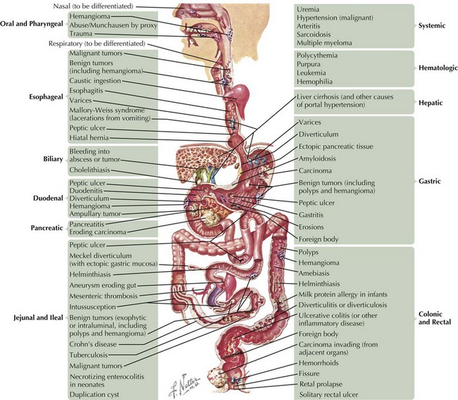
The causes of GI bleeding are diverse (Figure 108-1). Although the sheer number of causes may seem intimidating, grouping them into pathophysiologic categories aids in constructing a differential diagnosis. Note that underlying disorders of coagulation (e.g., hemophilias, vitamin K deficiency in neonates) and hepatic dysfunction (caused by the liver’s role in producing coagulation factors) can exacerbate any of these causes.
Direct Mechanical Injury
Blunt and penetrating trauma can initiate bleeding from any part of the GI tract. Ingestion of certain foreign bodies (e.g., coin batteries) may result in mucosal injury and even bowel perforation. Repeated vomiting or retching may create esophageal mucosal defects called Mallory-Weiss tears that can bleed. Surgical interventions, such as dental procedures and tonsillectomy, may result in blood loss that is swallowed and subsequently vomited.
Mucosal Erosion
The GI tract has a robust and sophisticated mucosal defense mechanism designed to prevent erosion. These protective elements include (1) the superficial “unmixed” layer of mucus, bicarbonate, and other factors that form a neutralizing barrier against acid, enzymatic, and abrasive injury; (2) the epithelial cells that generate this superficial layer; (3) continuous cell renewal coupled with (4) uninterrupted nutrient blood flow to the mucosa and (5) sensory innervation that optimizes this blood flow; and (6) endothelial production of prostaglandins and nitric oxide, which synergize to promote all of the aforementioned mechanisms. Disruption of any of these factors may predispose a given region of mucosa to erosion, local loss of vascular integrity, and resultant bleeding.
Drug-induced
A number of drugs can precipitate mucosal erosions, the most notorious being those from the nonsteroidal antiinflammatory drug (NSAID) class, which inhibit cyclooxygenase-mediated prostaglandin synthesis. Other culprits include corticosteroids, alcohol, caffeine, and nicotine. Chemotherapeutic agents, such as vincristine, methotrexate, and 5-fluorouracil, may cause GI bleeding via inflammation and mucosal erosion (termed mucositis) anywhere along the GI tract by inhibiting epithelial cell turnover, recruiting inflammatory cells, and making the mucosa more susceptible to infectious insults.
Inflammation
Disruption of mucosal integrity may occur in disorders that promote the recruitment of inflammatory cells, such as lymphocytes and neutrophils, which injure the epithelium by direct cell-to-cell contact or via secreted immunologic factors, such as cytokines. Examples include autoimmune enteropathy and eosinophilic gastroenteritis. Another important example is inflammatory bowel disease, a complex autoimmune disease that encompasses Crohn’s disease, which can cause full-thickness inflammation anywhere along the GI tract, and ulcerative colitis, which causes mucosal ulcerations in the colon (see Chapter 110). Children who have undergone bone marrow transplant may experience a form of rejection called graft-versus-host disease (GVHD). When it affects the GI tract, GVHD causes diarrhea, vomiting, fever, abdominal pain, and GI bleeding. Certain immunodeficiencies, such as common variable immunodeficiency, precipitate inflammation because of dysregulated immunity that leads to autoimmune responses (see Chapter 21).
Infection
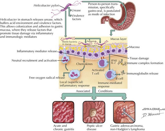
Infections disrupt mucosal integrity by direct cytotoxicity and/or by promoting inflammation. For example, herpes simplex virus is known to cause florid esophagitis, particularly in immunocompromised individuals, with nearly one-third of these patients experiencing an acute upper GI bleed. Additionally, infections may elicit specific peculiar responses. For example, cytomegalovirus is occasionally associated with massive gastric epithelial proliferation leading to a hypertrophic gastropathy called Menetrier’s disease. This causes protein wasting (including coagulation factors) from the leaky mucosa, as well as a higher risk of mucosal erosion and bleeding. Helicobacter pylori is perhaps the most infamous of the infectious causes of gastritis and duodenitis because of its chronicity, high prevalence (especially in certain populations of children, such as immigrants, the poor, and those attending daycare centers), its associations with peptic ulcers and adenocarcinoma of the stomach and duodenum, and its resistance to eradication (Figure 108-2).
Caustic Ingestions
Ingestion of acid substances causes a coagulation necrosis that results in ulcers and mucosal bleeding. Alkali ingestions can create significantly worse complications through liquefaction necrosis and mucosal sloughing, which can lead to severe ulceration, perforation, and eventual luminal stenosis.
Ischemia
Loss of blood flow to the bowel can lead to ischemic injury and necrosis. Examples of conditions in which this may occur are mesenteric arterial thrombosis; vascular malformations that create regions of suboptimal blood flow; and drug-induced bowel injury, as can occur with the use of certain chemotherapeutic agents such as vincristine and cisplatin. Depending on the severity of the occlusion, microscopic or frank bleeding may occur, typically accompanied by severe postprandial abdominal pain. Stress-related mucosal disease is a form of gastritis encountered in critically ill patients. The systemic hypotension frequently experienced in this population leads to local mucosal ischemia, causing erosions and subepithelial hemorrhage.
Infiltration
Although rare in children, tumors involving the GI tract, such as lymphoma, may promote bleeding by direct invasion and by inflammatory cells that may also damage the mucosa.
Hypersecretion
Several rare disorders may result in excessive or ectopic acid secretion. For example, gastrinomas are uncommon tumors arising in the stomach or pancreas that are a major cause of Zollinger-Ellison syndrome, in which pathologically elevated gastrin production promotes excess gastric acid secretion and the formation of peptic ulcers in almost all affected patients. Duplication cysts are developmental disorders in which ectopic gastric tissue within the cyst may cause inappropriate acid secretion in a relatively unprotected portion of the GI tract (potentially anywhere, most commonly the ileum), resulting in erosion and bleeding. They can also cause GI bleeding through other mechanisms, such as by acting as lead points for intussusception, with resultant ischemia, or through the rare development of a malignancy at the cyst site. Meckel’s diverticulum is another developmental disorder resulting from incomplete involution of the vitelline duct that can harbor ectopic gastric mucosa (features summarized by the rule of twos: 2% of the population, 2 feet from ileocecal valve, 2 inches long, most commonly presents around age 2 years, and boys are two times as likely to be affected).
Polyps
Polyps are mucosal projections into the intestinal lumen caused by disordered epithelial growth. Various spontaneous and familial polyposis entities exist. Regardless of the specific underlying process, they are a source of bleeding (generally painless) secondary to mechanical irritation or wholesale shearing by passing fecal material. In contrast to adults, the vast majority of polyps in children are juvenile polyps. These non-neoplastic polyps are typically composed of many cystic, dilated glands; copious inflammation; and surface erosions, the last of which is the cause of bleeding. Juvenile polyps have a peak incidence from 2 to 6 years of age, represent more than 90% of all pediatric polyp diagnoses, and are found in about 1% of school-age children.
Vascular Abnormalities
Congenital vascular defects may result in fragile vascular arrangements vulnerable to erosive and mechanical insults. Whereas hemangiomas represent proliferative vessel abnormalities that may regress, true malformations (including disorders such as arteriovenous malformations, blue rubber bleb nevus syndrome, and Osler-Rendu-Weber syndrome) are nonproliferative and do not regress. Processes that compromise the full thickness of bowel, such as Crohn’s disease and bowel surgery, may result in vessel-to-bowel fistulas associated with significant bleeding. Vasculitides, such as Henoch-Schönlein purpura, may result in occlusion of mesenteric vessels and intestinal ischemia (see Chapter 28).

Stay updated, free articles. Join our Telegram channel

Full access? Get Clinical Tree

