CHAPTER 56 Frequency, urgency and the painful bladder
Introduction
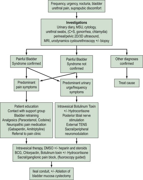
‘Cystitis’ is a term used for irritative urinary symptoms of frequency, urgency, suprapubic pain and dysuria. These symptoms of altered lower urinary tract sensation may be of recent onset (acute), longstanding (chronic) or recurrent. They may be caused by a variety of intravesical pathology such as infection, calculi, drug-induced inflammation, non-infective inflammatory processes due to local [e.g. painful bladder syndrome (PBS)] or systemic conditions (e.g. sarcoidosis), benign or malignant lower urinary tract tumours, or extravesical pelvic pathology such as pelvic masses (e.g. fibroids, endometriosis). Accurate diagnosis, appropriate intervention and therapy requires careful patient evaluation and a sound understanding of the differential causes (Figure 56.1).
![image](/wp-content/uploads/2016/06/B9780702031205000564_f1.jpg)
Figure 56.1 Management strategy for women presenting with intractable lower urinary tract symptoms. MSU, midstream urine sample; 2D, two dimensional; 3D, three dimensional; MRI, magnetic resonance imaging; TENS, transcutaneous electrical nerve stimulation; BCG, bacillus Calmette-Guérin; DMSO, dimethyl sulfoxide; C+S, culture and sensitivity.
Women with longstanding or recurrent chronic symptoms are often treated as recurrent bacterial cystitis. Often, it is only after a poor response to antibiotics or failure to culture uropathogens that an alternative diagnosis is considered. It is essential that these women have a thorough evaluation to exclude any serious underlying pathology (e.g. carcinoma) so that effective treatment can be commenced. Women with chronic irritative symptoms frequently have conditions such as PBS (formerly known as ‘interstitial cystitis’) or urethral syndrome. The pathogenesis for these conditions is poorly understood, and response to current therapy is often unsatisfactory.
Definitions
The Standardisation Sub-committee of the International Continence Society (ICS) published a terminology statement on lower urinary tract function in 2002 (Abrams et al 2002a,b). The majority of the following definitions are based on this statement.
Daytime frequency is the number of voids recorded during waking hours, and includes the last void before sleep and the first void after waking and rising in the morning. Increased daytime frequency is the complaint by the patient who considers that he/she voids too often during the day.
Nocturia is the complaint that the individual has to wake at night, once or more, to void. In other words, it is the number of voids recorded during a night’s sleep; each void is preceded and followed by sleep.
24-h frequency is the total number of daytime voids and episodes of nocturia during a 24-h period.
Bladder pain can be severe and frequently ill defined with radiation to the vagina and rectum. It is often aggravated by bladder distension, sexual intercourse, spicy foods, alcohol and caffeine, and relieved by voiding.
Dysuria is urethral pain during micturition and may be secondary to obvious pathology such as infection or a urethral diverticulum, or less clear causes such as atrophic urethritis or urethral syndrome.
Haematuria is an important symptom that requires urgent evaluation to exclude carcinoma.
PBS is the complaint of suprapubic pain related to bladder filling accompanied by other symptoms such as increased daytime and night-time frequency, in the absence of proven urinary infection or other pathology. The ICS believes this to be a preferable term to ‘interstitial cystitis’. Interstitial cystitis is a specific diagnosis requiring confirmation by the typical cystoscopic and histological features.
Urethral pain syndrome is recurrent episodic urethral pain, usually on voiding, with daytime frequency and nocturia in the absence of proven infection or other pathology.
Prevalence
A population-based study on the prevalence of lower urinary tract symptoms showed that the most commonly reported storage symptom in women is nocturia (54.5%). Urgency occurs in 12.8% and frequency in 7.4% of women. If nocturia is defined as two or more nocturnal micturitions per night, instead of one or more, the prevalence is decreased to 24% (Irwin et al 2006). PBS is a common condition in women, with reported prevalence of 1.7% in those aged under 65 years and 4% in those aged over 80 years. The vast majority of women with PBS have moderate or severe symptoms (Lifford and Curhan 2009).
Aetiology
Irritative bladder symptoms can be caused by a number of conditions originating within the lower urinary tract (Box 56.1). Infection and functional disorders, such as detrusor overactivity or voiding dysfunction, may cause urge symptoms and should be excluded. These conditions should be differentiated from more generalized systemic disorders (e.g. pregnancy, diabetes mellitus, renal disease), pelvic inflammatory disease or gynaecological surgery. A pelvic mass may cause urge-frequency symptoms due to bladder compression.
Box 56.1 Non-infective sensory disorders of the lower urinary tract
The aetiology of PBS is poorly understood. Several theories have been proposed over the years, including infection, immunological factors, leaky urothelium due to glycosaminoglycan deficiency, mast cell activation and altered neural function. Consensus is developing regarding epithelial dysfunction, mast cell activation and neurogenic inflammation; all part of a possible inflammatory response (Elgavish 2009).
Assessment
History
A detailed history should include specific questions regarding gynaecological and urinary symptoms. Frequency, urgency and nocturia in association with incontinence are likely to reflect underlying detrusor overactivity. In contrast, chronic severe urgency and bladder pain without incontinence are likely to be secondary to intravesical pathology such as infective or non-infective cystitis. A history of present and past medications [e.g. diuretics, tiaprofenic acid (Surgam) and cyclophosphamide], previous urinary tract infection (UTI) and pelvic surgery should be sought. Predisposing or exacerbating factors may include sexual intercourse (e.g. ‘honeymoon cystitis’), barrier contraception (e.g. diaphragm, condoms) and contact irritants (e.g. vaginal douches, spermicidal agents). More general irritant factors can include washing powder, soaps and gels.
Voiding dysfunction should be suspected with symptoms of hesitancy, poor urinary stream, straining and incomplete bladder emptying. Impaired bladder emptying due to detrusor underactivity or urethral obstruction may be idiopathic or secondary to neurological disease, pelvic surgery or uterovaginal prolapse.
Examination
Abdominal examination may reveal a palpable bladder secondary to urinary retention, pelvic mass or bladder tenderness from cystitis. Neurological examination with directed assessment of the S2–4 nerve roots, which innervate the bladder, should be performed. Vaginal examination using a Sims speculum allows visualization of the anterior and posterior walls, and may reveal genital prolapse, atrophy, inflammation or infection. Urethral inspection may reveal a mass, mucosal prolapse or caruncle. Paraurethral masses can be identified on bimanual examination by compressing the urethral and paraurethral tissues against the back of the pubic symphysis; expression of urethral pus suggests a urethral diverticulum.
Investigations
Urinary diary
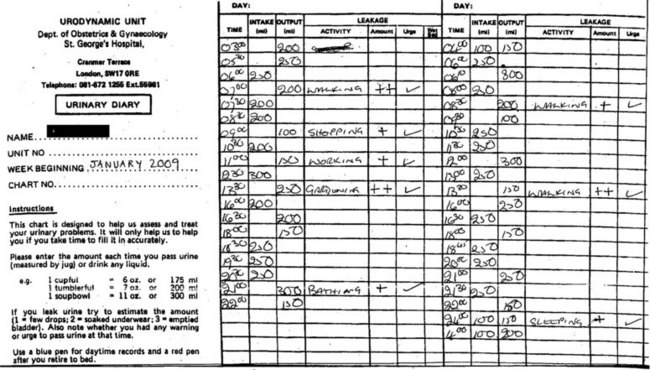
A detailed 3-day urinary frequency–volume diary is an important part of the initial and ongoing assessment (Figure 56.2). These diaries are more accurate than patient recall, allowing rapid and accurate diagnosis of urinary frequency, nocturia, estimation of voided volumes, functional bladder capacity, and assessment of episodes of urgency, pain and incontinence as well as their temporal relationship. A diary will also educate the patient regarding voiding habits, and is essential for bladder retraining.
Microbiology
Midstream urine sample (MSU) for microscopy, culture and sensitivity, and cytology will diagnose a UTI, confirming or excluding the presence of leukocytes, nitrites, red blood cells and any abnormal tumour cells shed from a bladder cancer. Isolated haematuria with a negative MSU may be caused by inflammatory cystitis or carcinoma, and is an indication for cystoscopy.
Urethral swabs are indicated where the MSU is negative and there is urethral tenderness, discharge or the patient is sexually active. In these cases, appropriate culture media should also be used for Chlamydia trachomatis and Neisseria gonorrhoeae. Other organisms commonly identified include coliforms and Staphylococcus saprophyticus. Tuberculosis is still prevalent, especially in developing countries, and should be considered with persistent sterile cystitis and pyuria. Diagnosis is based on a positive Lowernstein–Jensen culture using three early morning urine specimens.
Urodynamics
Urodynamic studies should be considered once infection has been excluded and symptoms have not responded to conservative treatment, especially if urinary incontinence is an associated problem. Uroflowmetry measures the voided volume, and maximum and average flow rates, and also records the voiding pattern. Filling cystometry gives information on bladder sensation, compliance and detrusor contractility, and may identify underlying stress incontinence, detrusor overactivity or voiding dysfunction. A diagnosis of bladder hypersensitivity is based on the following findings at urodynamics: stable bladder, capacity less than 350 ml and urgency at less than 150 ml. Both urodynamic assessment and a urinary diary will give an indication of functional bladder capacity and the severity of bladder hypersensitivity. Women with severe PBS/interstitial cystitis are frequently unable to hold more than 50–100 ml.
Imaging
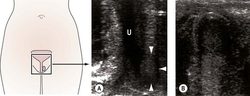
Imaging of the urinary tract can be performed using ultrasound or contrast radiography, either alone or synchronously with urodynamic assessment [videocystourethrography (VCU)]. VCU demonstrates voiding function and can identify urethral obstruction, a urethral diverticulum or external compression due to fibroids or prolapse. Haematuria in the absence of identifiable uropathogens, negative cystoscopy or recurrent UTI is an indication for assessment of the upper urinary tract using contrast intravenous urography or urinary tract ultrasound. Ultrasound can also determine bladder residual volume and bladder wall thickness, and identify any structural abnormalities, including urethral diverticulae (Figure 56.3) (Doumouchtsis et al 2008).
![image](/wp-content/uploads/2016/06/B9780702031205000564_f3.jpg)
Figure 56.3 Transperineal high-frequency ultrasound and corresponding line diagram demonstrating a large urethral diverticulum. U, urethra. (A) The arrows indicate the paraurethral lesion (diverticulum. (B) Enlarged ulltrasound image of the diverticulum in (A) demonstrating mixed reflective echogenic pattern.
![](https://freepngimg.com/download/social_media/63059-media-icons-telegram-twitter-blog-computer-social.png)
Stay updated, free articles. Join our Telegram channel
![](https://clinicalpub.com/wp-content/uploads/2023/09/256.png)
Full access? Get Clinical Tree
![](https://videdental.com/wp-content/uploads/2023/09/appstore.png)
![](https://videdental.com/wp-content/uploads/2023/09/google-play.png)