chapter 4 Evaluating the Newborn
Diagnostic Approach
If I had influence with the good fairy who is supposed to preside over the christening of all children, I should ask that her gift to each child in the world be a sense of wonder so indestructible that it would last throughout life.
Ideally, if you are going to care for a newborn, you should get to know the family before the child’s birth. This step is often taken by physicians in private practice but should be encouraged more widely. It makes antenatal care more comprehensive and helps the family care for the baby with as little anxiety as possible. Meeting the parents well before the delivery helps you become familiar with the prenatal history and makes you aware of important family issues. Your main sources of information should be the following:
The first-time mother of a 1-day-old baby boy who is being readied for discharge from the hospital has asked you to assess her baby. She is concerned that his color is poor and that he is not yet feeding well. When you ask about her specific concerns, she states that his hands and feet look blue when he is out to feed, that breastfeeding is not going well, and that he seems sleepy. The nurses have spent a lot of time with the mother trying to ease her concerns, but she is convinced that something is terribly wrong and that going home will put him in danger.
The infant, named Jonathan, was delivered vaginally at 38 weeks gestation following an uncomplicated pregnancy. When you arrive, both parents and the father’s sister are present and the mother is visibly upset and crying. The father voices the concern that Jonathan may have a heart condition underlying the color change and poor feeding, and he is refusing to allow his wife and baby to be discharged.
The experienced clinician will try to establish the root causes for the family’s concerns. For example, there may be another child with congenital heart disease with whom Jonathan is being compared. It also is important to allow the parents to voice their concerns about going home and to try to gain a better understanding about why the mother is so upset. A careful reexamination of the baby, preferably in the presence of the parents, is necessary.
There is a range of normal variation in peripheral cyanosis in the first few days after birth, particularly in situations in which the baby is exposed to cooler temperatures and when the limbs are in dependent positioning. Difficulties in establishing breastfeeding also are not uncommon, and most infants, after initial periods of wakefulness and good attempts at feeding, will experience a period of increased sleeping and less vigorous feeding. When infants and mothers are first learning to breastfeed, it is important that attention be paid to thermoregulation and positioning. Peripheral cyanosis, which is most often benign, is not uncommonly interpreted by brand-new parents as a worrisome sign and results in excessive anxiety and concern.
This chapter describes the usual activity of a newborn infant with tips on how to distinguish normal neurologic signs of tone and activity. A clear description of the cardiovascular examination is given, including some useful information about when to become concerned about murmurs or change in pulses.
Obtaining the History
Table 4–1 lists the important issues to ask about when obtaining any antenatal history.
TABLE 4–1 Significant issues to ask about when taking the History for a Newborn
Mother’s past pregnancies and their outcome | No. of pregnancies |
Stillbirths | |
Abortions | |
Neonatal deaths | |
Cesarean sections | |
Specific concerns that parents may have about current pregnancy because of past experiences | |
Preexisting systemic illness in mother and maternal medications | Hypertension |
Depression | |
Diabetes | |
Seizure disorder | |
Thyroid disease | |
Cardiac disease | |
Metabolic disorder (phenylketonuria) | |
Genetic history | History of inherited disorders |
Consanguinity | |
Unexplained neonatal deaths in the family | |
History of current pregnancy | Date of last menstrual period |
Use of assisted reproductive technologies or fertility treatments | |
Estimated date of conception by dates (and by ultrasonography if early ultrasonogram [9–13 weeks] available) | |
Results of ultrasonography, amniocentesis, cordocentesis, chorionic villus sampling | |
Pregnancy-induced hypertension, gestational diabetes | |
Note maternal weight gain and blood pressure, fetal growth, blood type | |
History of alcohol use, drug use (prescribed or illicit), cigarette use | |
Group B Streptococcus status (if known) | |
History of maternal surgery during pregnancy | |
Concerns about placenta (e.g., placenta previa or thickening) | |
Use of magnesium sulfate, betamethasone | |
Current labor and delivery | Induced or spontaneous labor (if induced, why?) |
Time of rupture of membranes and quality of amniotic fluid (bloody, meconium-stained) | |
Length of second stage | |
Use of medications (analgesics and time prior to delivery) | |
Intrapartum fever and antibiotics | |
History of fetal distress | |
Presentation (vertex, breech, transverse) | |
Vaginal or cesarean delivery (if cesarean, why?) | |
Use of forceps or vacuum extraction | |
Adaptation to extrauterine life | Apgar scores |
Resuscitation needed? If so, what and for how long? | |
Need for naloxone |
Approach to Physical Examinations of the Newborn: When, Why, and How
The first examination
Every newborn infant should be examined at least twice during the first few days of life. An additional examination after discharge but within the first week is recommended, particularly if the mother and infant are discharged from the hospital prior to 48 hours postnatally. If possible, perform the first assessment in the hospital’s delivery area to (1) identify any obvious major and minor congenital malformations, (2) assess gestational age, nutritional status, and vigor, and (3) determine how well the baby handles the transition from intrauterine to extrauterine life.
Most babies make the major physiologic adaptations of this transition smoothly. The few who do not adapt normally—particularly those born prematurely or after perinatal stress or asphyxia—will need help to adapt successfully.
The 1-minute Apgar score should not be used to determine if resuscitation should be started. Resuscitation, if needed, should start immediately.
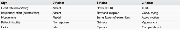
The method used almost universally in delivery rooms to evaluate the central nervous system (CNS) status and general adaptation of the neonate to extrauterine life is the Apgar score. This scoring system, summarized in Table 4–2, evaluates the baby in five different respects: heart rate, respiration, color, muscle tone, and reflex irritability (response to stimulation). These signs are usually evaluated at 1 minute and 5 minutes after birth. The Apgar score is the total of the baby’s scores for each of the five signs. To be consistent, have the Apgar score recorded by an experienced clinical observer whose principal responsibility is caring for and evaluating the baby. Because inter-observer variation in scoring may be considerable, experience is essential. Evaluate the signs at exactly 1 minute and 5 minutes to establish the score, especially in relation to muscle tone and reflex irritability. The latter is usually elicited by firmly stroking the sole of the foot; the appropriate response to this stimulation is a vigorous cry.
Recording the Apgar score serves two purposes. First, it ensures a careful evaluation of the baby during the immediate newborn period. Second, it helps to determine the presence and level of CNS depression and whether there is a need to continue resuscitation. In babies who need resuscitation, serial scores measured at 1, 2, 5, and even 10 minutes furnish a semi-quantitative method of recording recovery. The relationship between low Apgar scores and neurologic abnormality later in infancy is not reliable.
The Apgar scoring system produces more than just a number. The presence or absence of individual signs should tell you not just that an infant had an “Apgar 6” but that this score was assigned because certain functions were absent or suboptimal. For example, a baby with congenital neuromuscular disease may score well for heart rate, respiratory effort, and color but poorly for muscle tone and reflex irritability. A preterm infant scores lower than a full-term baby because an immature baby’s muscle tone is always less than that of a normal-term infant.
The accurate diagnosis of perinatal asphyxia is not easy and requires not only evaluation of the clinical condition at birth but also evaluation of umbilical cord arterial blood gases and subsequent clinical neurologic sequelae in the newborn period (hypoxic-ischemic encephalopathy). Low Apgar scores may be caused by many conditions affecting the baby, such as sepsis, neuromuscular disorders, and perinatal asphyxia.
The second examination
The best place to carry out the second physical examination is in the mother’s room, preferably with both parents present. Having the parents there makes it possible to obtain further history details, complete the initial physical examination, answer questions on the spot, demonstrate and discuss normal variations or abnormalities discovered during the physical examination, and offer some valuable anticipatory guidance. This second, more thorough examination should be conducted within the first 12 hours of life. It is designed to identify congenital malformations, ascertain that adaptation to extrauterine life is proceeding normally, and detect any prenatally or perinatally acquired illnesses. The sooner it is performed, the happier the parents will be.
The third examination
The main purposes of the third examination are to discover postnatally acquired problems, such as infection and excessive jaundice, and to detect any malformations that were not apparent at the first or second examination, such as some forms of congenital heart disease whose murmurs are not audible on the first day of life. In past years, the third examination often was performed at 3 or 4 days of age. The current trend toward early discharge of mothers and infants from the hospital means that discharge may occur as early as 24 hours after birth, and discharge is common by 48 hours after a normal vaginal delivery. Thus the third examination often takes place after the mother has been discharged from the hospital. Early hospital discharge guidelines now recommend assessment of a newborn by a health care provider within 72 hours of discharge. However, it is important that this third examination and assessment of the baby’s condition be carried out within the first week of life. The third examination may be performed by a physician or by an experienced nurse or midwife and may take place in the hospital, in the home, or at a clinic or office.
Initially, many of us learned how to perform a physical examination with cooperative adult patients, in whom it was easy to start at the top of the head and work downward in a systematic cephalocaudal manner. Because infants are not always cooperative, you must be prepared to examine a newborn infant thoroughly but to keep the sequence of procedures flexible. For example, if the baby is resting quietly, first listen to the heart and chest, then palpate the abdomen. Carry out other parts of the examinations when the baby is active, moving, or even crying, but be sure to examine all systems and record the findings systematically.
Clinical Observations and What They Mean
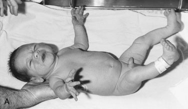
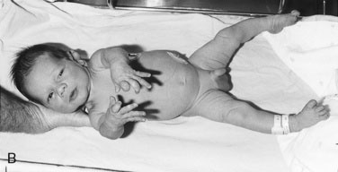
Encourage the parents to participate as much as possible in the examination by undressing the baby, holding the baby on the lap or the bed, and providing a stabilizing finger for the baby to grasp (Fig. 4–1). The first part of the examination is the most important. Do not touch the baby except to remove all clothing gently. You may wish to leave the diaper on until you are ready to examine the lower portion of the baby. The baby should be undressed, warm, and well illuminated and should be helped to feel stable and secure on the examining surface. Improving the baby’s stability and relaxation by letting him or her grasp the parent’s or your finger allows an immediate appreciation of the strength of the baby’s reflex grasp and reduces the tendency for instinctive startle reflexes that occur when the baby feels unstable and rolls on the examining surface.

FIGURE 4–1 The newborn feels more stable and tends to relax if you allow the infant to hold your hand during the examination.
The ideal time to examine a newborn is a couple of hours after a feeding, when the baby may not be too deeply asleep, as babies often are just after a feeding, nor awake and screaming, as they often are just before a feeding. Stand back and take a long hard look at the infant; educated observation often reveals far more than touching, poking, and kneading. While observing the newborn, answer the following questions:

FIGURE 4–2 Cutis marmorata (“marble skin”) can occur as a normal phenomenon when the baby is exposed.
Look for hematomas, hemangiomas, ecchymoses, and petechiae. The last can be associated with increased intravascular pressure during delivery, thrombocytopenia from the presence of platelet antibodies, or congenital infections. Cutaneous hemangiomas are often absent or minimally apparent at birth but develop and grow during the first weeks or months of life, only to regress spontaneously later. Ecchymoses may indicate more than usual trauma or an underlying coagulation disorder.
You can perform much of the newborn’s neurologic examination without touching the baby. Do all four extremities move equally and maintain a general posture of flexion, with alternating flexion and extension movements? The eyes should move if they are open, and the baby may exhibit sucking and tongue movements, yawning, facial grimacing, and spontaneous startle responses. The four most important parts of the neurologic assessment are:
A baby’s level of alertness varies with the time of day and the time of last feeding; normal newborns spend about 20 out of 24 hours asleep. Visual alertness is temporarily limited if erythromycin ointment has been instilled in the eyes as prophylaxis for gonococcal ophthalmia.
Listening To the Cry
Babies normally cry when they are uncomfortable, disturbed, or hungry. Train yourself to listen carefully to infant cries, because there is much to be learned from them. The normal cry should be strong; hoarseness, weakness, or an unusually high-pitched or low-pitched cry may indicate laryngeal or neurologic abnormalities. Repetitive inconsolable crying also is abnormal. An experienced nurse or mother can quickly tell you whether a particular baby’s cry is normal. Sometimes the nurse or mother is the first to suspect a disorder simply on the basis of an unusual cry, such as the high-pitched “cerebral” cry; the low throaty cry of congenital hypothyroidism; the characteristic catlike cry of the cri du chat syndrome; or the weak, poorly sustained cry of the infant who is sick.
Muscle Tone
By carefully examining many babies, you will become familiar with the differences between normal, increased, and diminished muscle tone in newborns. The differences cannot be described in words. Only by making it a regular practice to flex and extend babies’ arms and legs repeatedly during every examination of newborns can you develop an instinctive “feel” for normal, increased, and decreased muscle tone. When a newborn baby is pulled to the sitting position from the supine, the head lags behind the trunk; a healthy full-term baby makes some effort to bring the head in line with the chest. Once lifted to a sitting position, a full-term infant can keep the head upright for several seconds.
Primitive Reflexes
Many so-called primitive reflexes can be elicited that may already have been observed during the earlier part of the examination.
Asymmetric tonic neck response
Assess for the asymmetric tonic neck response by turning the baby’s head from midline to one side and noting the arm’s gradual extension on the side to which the head is turned, along with flexion of the other arm. The position resembles the classic fencing or boxing posture. Because of this reflex, it is important to ensure that the baby’s head is midline before you elicit any of the other responses.
Moro reflex
Newborns often demonstrate the Moro, startle, or embrace reflex spontaneously when they are suddenly moved, exposed to a sudden loud noise, given the feeling of falling, or feel unstable on a flat surface. You can most reliably elicit this reflex by allowing the infant’s head to drop (while supporting it gently) below the level of the rest of the body while the baby is in a reclining position. The baby extends the arms suddenly and rapidly with the hands open, then brings them back together more slowly in a movement that looks like an embrace. The initial rapid movement may be accompanied by a grimace or cry (Fig. 4–3). Symmetry of movement is important. Weakness of one arm (e.g., in an infant with brachial plexus injury) results in an asymmetric Moro reflex.


FIGURE 4–3 Induce the Moro or “embrace” reflex by giving the infant the feeling of falling. The response begins with a sudden extension of the arms and legs (A), which is followed by a slower embracing movement in which the arms are brought together (B). The Moro reflex is sometimes accompanied by a cry.
Palmar grasp
Find the palmar grasp by placing your index fingers in each of the baby’s open hands; the baby’s fingers should close around your fingers with strength enough to lift the baby’s body part of the way off the bed or table (Fig. 4–4).
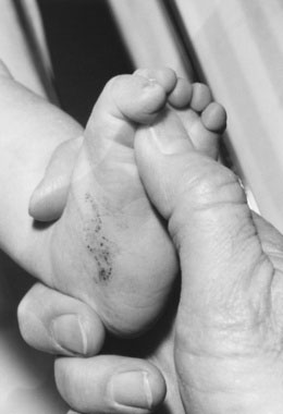
Plantar grasp
A strong plantar grasp usually can be demonstrated if you place your thumb on the sole of the baby’s foot in the space under the toes (Fig. 4–5).
Placing and propulsive (stepping and walking) reflexes
Holding the baby upright, touch the dorsum of the feet to the bed or a table, and it will seem as if the baby is trying to walk. Parents are invariably fascinated and delighted by the demonstration that their newborn baby can “walk.”
If there is any doubt about the integrity of the baby’s CNS, either because of the history (difficult labor, delivery, traumatic procedures, or birth asphyxia) or because of associated physical findings, such as an abnormal cry, major deformities, or malformations, a more complete neurologic examination should be conducted (see Chapter 13).
Weighing and Measuring
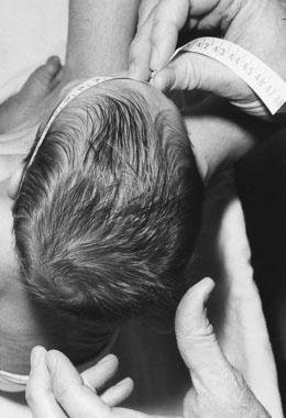
For the physician’s and the parents’ records, it is important to (1) measure the baby’s head circumference, length, and weight accurately and (2) assess the nutritional status. Measure the head circumference, preferably with a disposable tape measure, around the largest occipital-frontal diameter across the forehead, just above the eyes, and over the most prominent part of the occiput (Fig. 4–6). Head circumference normally measures 33 to 37 cm in a full-term infant. If a baby’s head circumference measures slightly above or below the normal for age, check the parents’ head circumferences before “pushing the panic button.” Benign familial megalencephaly, a normal variant, is the most common cause of a larger than average head. The condition can be verified by checking the head circumference of both parents. Megalencephaly is usually handed down by the father.

Stay updated, free articles. Join our Telegram channel

Full access? Get Clinical Tree

