Evaluating Gastrointestinal Symptoms

chapter 11 Evaluating Gastrointestinal Symptoms


Gastrointestinal (GI) symptoms are common in children, and evaluating their complaints can be challenging. This challenge is compounded by the fact that younger patients cannot communicate or localize their symptoms.


No amount of reading can substitute for hands-on practice with real patients. Paracelsus once remarked, “The art of medicine cannot be inherited, nor can it be copied from books.” This chapter offers clinical tips on how to evaluate GI symptoms in infants and children and how to perform a competent examination and arrive at a diagnosis. The focus is on evaluating symptoms and signs, not on investigations or management. Seven common symptoms, representing the bulk of GI complaints in children, are discussed. Of all the possible diseases causing these symptoms, only the more common disorders will be addressed.


General Appearance


Begin your examination by assessing the child’s growth, that is, weight, height/length, body mass index, and head circumference (in infants and toddlers), and plot these values on the appropriate growth charts. “Eyeballing” is not good enough. Sometimes more detailed anthropometric measurements may be required, including mid-arm circumference and skin fold thickness. Sexual maturation should be assessed in older children and adolescents. Recent loss of fat tissue may lead to loose, wrinkled skin, especially notable in the groin and as flattening of the buttocks. Peripheral edema may suggest hypoalbuminemia from a protein-losing enteropathy and/or poor nutritional intake.


Poor growth is an important feature of many organic GI diseases and usually rules out a functional disorder. Growth failure can be the result of inadequate nutrient intake (as in Crohn’s disease) or malabsorption (as in cystic fibrosis and celiac disease). Adequate growth is reassuring but does not necessarily rule out an organic GI disorder.


The child’s general appearance and behavior can provide important clues to the cause and severity of the problem. Anorexia and pain/irritability are common in many patients with acute and chronic GI diseases. Sudden, marked irritability with intense crying in an infant, especially when intermittent, should raise the possibility of intussusception. An ill child is often withdrawn, irritable, maintains poor eye contact, and is not interactive or interested in the surroundings. These signs are especially significant in infants and toddlers whose verbal communication may be limited. A toddler with fever and vomiting who is running around the emergency department with a bottle of juice is unlikely to have an acute abdomen.


Examination of the Abdomen


Examine the abdomen systematically. The child should be lying flat and supine, arms by the side, and feet together. The abdominal muscles are most relaxed in this position. The knees can be flexed if this position helps the youngster feel more comfortable. A pillow under the head also may be used. Infants and toddlers who may resist lying down can be examined while sitting on the parent’s lap. It is futile to attempt abdominal palpation in a youngster who is upset or crying. Try to gain the child’s cooperation. Otherwise, defer the examination and try again when the child is quiet or asleep. Infants are best examined after feeding.


The abdomen should be fully exposed. Older children and adolescents should be in a gown, or a sheet can be used to cover the body. Examination of the inguinal region, external genitalia, and perianal area (in select cases) is part of the abdominal examination, especially in infants who cannot localize their symptoms. For example, an infant presenting with acute onset of irritability, poor feeding, and vomiting may have a strangulated inguinal hernia. Examination of external genitalia is especially important in male patients because a variety of testicular and penile lesions can occur. A toddler with testicular torsion may present with severe abdominal pain and be unable to localize the pain. Often physicians are hesitant to examine the external genitalia in children, especially if the complaint is not directly linked to that region. However, serious diagnoses can be missed if the relevant areas are not specifically examined.


When examining a child with acute abdominal pain, it often is necessary to decide whether the child has a problem requiring surgery (i.e., an acute abdomen). Surgical disorders often present with a shorter history, and the most common cause of an acute abdomen in children is appendicitis. Disorders presenting with acute abdomen are discussed in more detail in Chapter 12.


Abdominal examination should follow the classical sequence, that is, inspection, palpation, percussion, and auscultation. The basic rule is, “Look first, touch later.” Some adult gastroenterologists recommend that auscultation be performed before palpation or percussion on the assumption that bowel sounds induced by palpation may mask vascular bruits. However, for some young children, the stethoscope can be intimidating if used at the beginning of the abdominal examination.


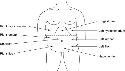
To describe the location of any abnormality, it helps to divide the abdomen into four quadrants with a horizontal line through the umbilicus and a vertical line from the xiphoid process to the symphysis pubis through the umbilicus. For infants and younger children with smaller abdomens, this division should suffice. For older children and adolescents, the abdomen can be divided into nine regions by including two additional vertical lines from the mid-clavicular area to the mid-inguinal point and two horizontal lines through the subcostal margins and the anterior iliac crests (Figure 11–1).


Inspection


Simply looking at the abdomen can provide important clues to the underlying diagnosis. Is it flat, distended, or scaphoid? Distention can be caused by air in the intestine, fluid in the abdominal cavity (ascites), or solid organ enlargement. Healthy toddlers often have a normally protuberant abdomen as a result of physiological increase in lumbar lordosis. Do not mistake this appearance for pathologic distention. Abdominal distention is an important sign of intestinal obstruction. The lower the obstruction in the gut, the more marked the distention. Also, associated symptoms will be present, including vomiting and abdominal pain/irritability. Peristalsis may be visible through the abdominal wall in patients with intestinal obstruction. This peristalsis is most commonly observed in the upper abdomen in infants with hypertrophic pyloric stenosis. A depressed (scaphoid) abdomen in a newborn suggests a diaphragmatic hernia with some abdominal organs sitting in the chest cavity.


Umbilical hernias are common in infants. In some ethnic groups, they are virtually universal. Umbilical hernias are reducible and, although they are sometimes quite large in size, they typically resolve without treatment during the first few years of life. Slight separation of the rectus abdominis muscle (diastasis recti) is normal in children, especially in premature infants.


Scar(s) on the abdominal wall will confirm previous surgical intervention. Information regarding previous operations usually is available in the history; however, operations in the remote past may be forgotten. With laparoscopy now being the preferred technique for most abdominal operations, scars may be minimal and difficult to locate.


The abdominal wall normally moves with respiration, especially in infants. Loss of this movement secondary to pain can be an important indicator of abdominal inflammation, such as peritonitis.


Abdominal symmetry is best assessed by inspection from the foot of the bed. Asymmetry can result from an intraabdominal mass. Bulging of the flanks can occur in patients with ascites.


Examine the inguinal area for swelling. Indirect inguinal hernias are common in children and always require surgical correction. A reducible hernia may not be obvious in the supine position. Older children should be examined standing up. Increasing the intraabdominal pressure by coughing or straining makes the swelling more obvious. Small, nontender, soft, mobile inguinal lymph nodes are common in children. They can be bilateral and often are benign. Check for abnormal lymphadenopathy elsewhere (e.g., the axilla).


Examine the back, because diseases of the spine may present with abdominal symptoms. A tuft of hair in the midline often is a clue to an underlying congenital spinal abnormality (e.g., diastematomyelia) in a child with chronic constipation.


Palpation


A relaxed abdomen is essential for palpation. Your hands should be warm and your touch gentle. The child should not laugh or giggle during the examination. The child’s attention should be diverted by conversation or by asking him or her to look at something on the ceiling.


Use superficial palpation to detect areas of tenderness. Watch the child’s face for any expression of discomfort. If the child presents with complaints in a specific area, begin palpation away from that area, then move gradually toward the site of pain. Check for guarding and rebound tenderness whenever you discover an area of tenderness on palpation. Deep palpation should be followed by quick release of pressure. If the patient has more pain when the hand is released, rebound tenderness is present. Rebound tenderness is an important sign of peritoneal irritation but can be difficult to demonstrate in very young children.


Deeper palpation should follow to detect any masses or organ enlargement. By far, the most common abdominal mass in children is retained stool. In newborns, a large mass is usually of renal origin. Fecal masses can be felt in the hypogastrium and left lower quadrant. Sometimes their massive size can be alarming. Fecal masses often are indentable and nontender. Ovaries normally are not palpable in healthy girls. The possibility of pregnancy should always be considered in any pubertal female with abdominal symptoms. You can get your hand above an ovarian or uterine mass, confirming its pelvic origin.


The liver, spleen, and kidneys should always be palpated.


Liver


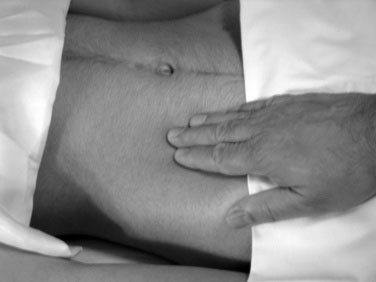
Always begin your palpation in the right iliac fossa so as not to miss the edge of a massively enlarged liver. The axis of your hand should be directed toward the right costal margin and at right angles to it. Rest your fingertips on the abdomen and gently depress them intermittently, without poking (Fig. 11–2). The child’s respiratory excursions will bring the liver down to make contact with the fingers (you don’t feel it; it feels you). One or both hands (one on top of another) may be used. Advance your fingers upward toward the costal margin in 1- to 2-cm increments.


If you can palpate the liver edge, confirm its edge by percussion. Record any hepatomegaly as centimeters below the costal margin, not in finger breadths. Palpate the epigastrium, because isolated enlargement of the left lobe (because of a tumor or cyst, for example) may be present.


The liver may be palpable either because it is enlarged (hepatomegaly) or because it has been displaced downward by thoracic deformities or by flattening of the diaphragm, for example, by a large, right-sided pleural effusion or lung hyperinflation (e.g., bronchiolitis). Measuring the liver span gives a good estimate of its size. It also helps distinguish between enlargement and displacement. The lower border of the liver should be marked. Start percussion for the upper border from the third intercostal space and move down one intercostal space at a time. The finger should be in the intercostal space parallel to the ribs. The distance between the upper and lower borders (span) should be measured in the mid-clavicular line. Use of the “scratch test” to define the lower border of a nonpalpable liver is not useful in children.


The liver often is palpable 1 to 2 cm below the right costal margin in normal infants and toddlers. Its edge is usually soft, and it moves downward with respiration. The liver edge is often palpable in healthy children and adolescents. A nonpalpable liver does not rule out hepatomegaly but certainly reduces its likelihood.


Liver span in children is related to age and body weight, with a considerable range. As a rough guide, the liver span ranges from 6 to 12 cm in children 6 to 12 years of age. In infants up to a year of age it is 6 to 10 cm. A palpable liver with a normal span implies displacement rather than enlargement. Measuring span is important when the liver is palpable. In a patient with a shrunken, cirrhotic, nonpalpable liver, span would be difficult to measure because the lower border cannot be identified easily. Estimation of liver span caries considerable observer error and can be difficult in female adolescents, in whom breast tissue can make percussion of the upper border unreliable. Ultrasonography gives a good estimate of liver size.


The liver texture can give clues to the cause of hepatomegaly. An enlarged, soft liver is due to inflammation or congestion. A firm liver may represent fibrosis, metabolic disease, or primary or secondary malignancy.


Hepatomegaly can occur from many different causes, including:


Spleen


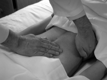
The spleen tip is palpable in about 10% of healthy children and often is palpable in newborns. In infants, the spleen enlarges downward toward the left lower quadrant, whereas in older children enlargement is toward the right lower quadrant. Palpation should begin in the right iliac fossa so as not to miss a very large spleen (Fig. 11–3). Be gentle, because an enlarged spleen could rupture if too much force is applied. Size should be recorded in centimeters below the left costal margin.


If the spleen is not palpable with the child supine, turn the child into the right lateral decubitus position. With your left hand lifting the lowermost portion of the left rib cage and flank anteriorly, your right hand should be used to feel the spleen tip on inspiration. In subclinical splenomegaly, other provocative maneuvers may be tried. Castel sign involves percussing the lowest intercostal space in the left anterior axillary line. Normally this area is resonant and should stay so during inspiration. An older child can be asked to take a deep breath while percussion is continued. A change from resonance to dullness suggests splenic enlargement.


Occasionally, it may be difficult to differentiate splenomegaly from a massively enlarged kidney. With an enlarged spleen, one cannot get above it, a notch may be felt along its medial border, and the percussion note over it will be dull.


Is a palpable spleen abnormal in a child? It depends on the clinical context. An enlarged spleen is present in many patients with chronic hemolytic disorders. Sickle cell anemia is an exception because the spleen atrophies over time as a result of repeated infarctions. Splenomegaly is an important finding in portal hypertension from any cause. If an enlarged spleen is present along with hepatomegaly, consider infections, infiltrative disorders, or storage disorders.


Kidneys


Kidneys are rarely palpable in healthy children. A kidney may be enlarged because of an obstruction (hydronephrosis), an infection, a cyst, or a tumor. Bilateral renal enlargement suggests either polycystic kidneys or bilateral obstructive uropathy due to posterior urethral valves.


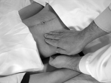
Kidneys are retroperitoneal and deep and are best palpated bimanually. With the child supine, place one hand in the renal (costovertebral) angle, beneath the twelfth rib and just lateral to the erector spinae (Fig. 11–4). Place your other hand anteriorly, just lateral to the rectus abdominis, in line with and parallel to the first hand. Ask the child to take a deep breath. Immediately at the end of inspiration, press your front hand firmly back against the other hand. A quick upward movement of the fingers of the hand in the renal angle is made (ballottement), and the kidney is trapped between the two hands. Unless the kidney is massively enlarged, successful renal palpation requires considerable experience. The percussion note over the kidney will be resonant because of overlying intestine, which helps differentiate it from the spleen or liver.


Renal angle tenderness often is associated with renal disease. The heel of the closed fist is used to strike the patient gently over the renal angles to check for tenderness.


Rectal Examination


There are few indications for doing a rectal examination in children. It is rarely required in children with constipation because the diagnosis often can be made from the history and abdominal palpation. Perianal inspection may be indicated in cases of rectal bleeding and suspected Crohn’s disease. Children do not get hemorrhoids.


If a rectal examination must be performed, it should be done systematically. Consent from the parents and assent from the child is essential. Some institutions may require a written consent for a rectal examination. The procedure should be explained in detail. If the child refuses, do not insist. The examination should not be attempted in an uncooperative child who is crying and struggling; not only would it be traumatic, but it would be virtually impossible to do an adequate digital rectal examination under such circumstances. The privacy of even the youngest children should be respected. A nurse should be present to assist in proper draping of the patient. The parent stands on the head side of the bed to provide comfort.


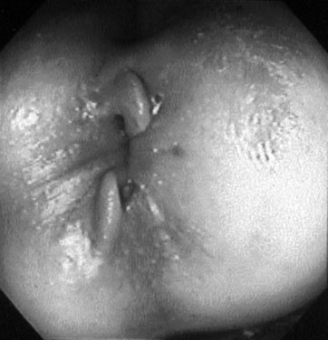
In older children, a rectal examination is best performed with the child in the left lateral position with the spine and knees fully flexed. Infants and younger children can be examined in a knee-chest position. The examination should be conducted systematically. Presence of stool in the underwear implies fecal incontinence, most likely from encopresis. Examine the perianal area for excoriations, skin tags, or fistulae. Sentinel skin tag(s) overlying a chronic anal fissure in older children are pathognomonic of Crohn’s disease (Fig. 11–5). The buttocks should be gently spread to look for any anal fissure. Acute anal fissures occur from passage of large, very hard stools. Digital rectal examination in such cases is painful and unnecessary. Acute anal fissures often are difficult to view because there is considerable spasm of the anal sphincters, and the child does not relax enough for good visualization. The diagnosis is best made by history.


Next, check the anal reflex (wink). Stroking the perianal skin close to the anal verge causes contraction of the anal sphincter, similar to the cremasteric reflex. Absence of the anal wink should raise suspicion of an underlying neurologic impairment. A well-lubricated forefinger is then introduced in the rectum. In infants, the little finger should suffice. The anal squeeze is normally felt. A patulous anus is seen in patients with encopresis or neurologic disorders. Is the rectal cavity empty or full of stool? Is the stool hard? Is any other mass present? The normal rectum should be empty with a smooth wall. In functional fecal retention, the rectum contains a large mass (fecaloma), which often can be indented. An empty rectum in the absence of recent defecation and a background history of chronic constipation should raise suspicion of Hirschsprung disease. Finally, the finger should be withdrawn to look for the presence of blood.


Percussion


Percussion of the abdomen helps to detect free fluid and to determine whether distention, if present, is due to gas, a solid tumor, or ascites. Gas produces a resonant note. In liver and spleen enlargement, the degree of organomegaly can be confirmed by percussing the lower edge. The measurement should be recorded in centimeters below the costal margin in the midclavicular line.


When percussing for organomegaly, the resting finger should be parallel to the edge of the organ being examined. Contact with the skin should be gentle. Percuss by gently tapping the middle finger of the right hand against the finger in contact with the skin. The movement of the hand should be at the wrist, not the elbow. A dull note implies that the underlying mass is solid or filled with fluid.


Shifting dullness and fluid thrill (wave) are clinical signs that confirm ascites. Free fluid will flow to the dependent part of the abdominal cavity, while the gut filled with air will float in the middle. If the child is supine, the fluid collects posteriorly and in the flanks. The percussion note, therefore, will be resonant in the center and dull in the flanks. Starting in the middle, the percussion is carried laterally toward the flank, where the note will become dull. Ask the child to roll over to the opposite side. The previous area of flank dullness now becomes resonant as the fluid moves to the dependent part of the abdominal cavity. The same technique can be used for either flank.


To elicit a fluid thrill, the child should lie supine. Place one hand flat on the flank and tap or flick the opposite flank briskly with the other hand. If fluid is present, a wave is felt by the former hand. To dampen any wave that may pass through the fat in the anterior abdominal wall, a third hand (that of the patient or an assistant) is placed vertically in the midabdomen. Use of the third hand is rarely needed in children unless the child is obese.


Demonstrating shifting dullness is helpful when a small amount of free fluid is present in the abdomen. With massive ascites, a fluid thrill is present but shifting dullness may not be demonstrable, because there is little room in the abdominal cavity for the fluid to shift. In infants and young children, a small amount of ascites can be difficult to confirm by clinical examination, and abdominal ultrasonography may be required.


Ascites most commonly occurs in patients with cirrhosis of the liver, but it also may occur in patients with right heart failure, hypoalbuminemia, lymphatic obstruction, and primary abdominal malignancies.

< div class='tao-gold-member'>

Stay updated, free articles. Join our Telegram channel

Jul 3, 2016 | Posted by in PEDIATRICS | Comments Off on Evaluating Gastrointestinal Symptoms

Full access? Get Clinical Tree

Get Clinical Tree app for offline access