CHAPTER 34 Epidemiology of gynaecological cancer
General Overview
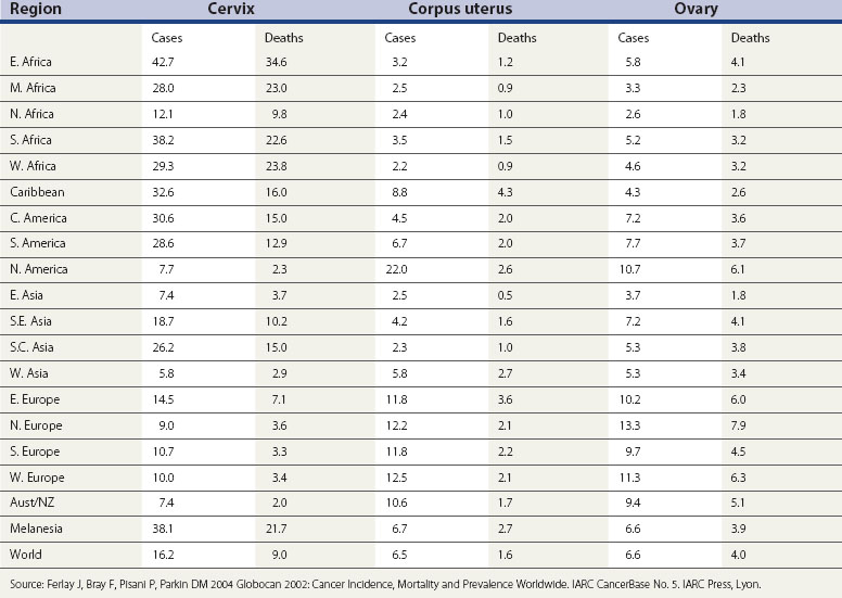
Globally, breast cancer and cancer of the cervix are the two most common female malignancies. However, both ovarian and endometrial cancer rank in the top 10 female malignancies. Cervical cancer is most common in developing countries, while cancer of the uterine corpus and ovarian cancer have higher incidence in industrialized countries. Indeed, in the presence of cervical screening, these cancers are more common than cervical cancer in most Western countries. Table 34.1 provides an overview of age-standardized incidence and mortality rates of these three gynaecological cancers around the world. Other gynaecological cancers (i.e. cancers of the vulva and vagina) are relatively rare in all parts of the world. The age-standardized rates are less than one per 100,000 in most countries (Curado et al 2007).
Table 34.1 Age-standardized rates (per 100,000 women-years) for cancer of the cervix, corpus uterus and ovary (world standard population)

The variation in the rates of gynaecological cancers around the world is enormous (Table 34.1). Cervical cancer rates in East Africa are 12 times greater than in West Asia (the Middle East). The rates for cancer of the uterine corpus are nearly 10 times higher in North America than in West Africa, and ovarian cancer is two and a half times more common in Northern Europe than in Middle Africa. Some, but certainly not all, of these variations can be explained in terms of differences in lifestyle and public health interventions.
Cervical Cancer
The cervical epithelium is composed of two distinct cell types. The ectocervix is covered by non-keratinized squamous cells similar to those of the lining of the vagina. The endocervical canal is covered by columnar cells of the same origin as those of the endometrium. Cervical cancers initiate in the region where these two cell types meet — the squamo-columnar junction. There are three main types of cervical cancer (squamous, adenocarcinoma and adenosquamous carcinoma), with squamous cell carcinoma being the most common. It used to be said that this accounted for approximately 90% of all cases of cervical cancer. However, more recent data show that the proportion of cervical cancer that is adenocarcinoma or adenosquamous carcinoma has doubled, particularly in younger women. Squamous cell carcinoma now only accounts for approximately 75% of all cases of cervical cancer. The reason for the increasing proportion of adenocarcinoma seems to be three-fold: adenocarcinoma really is becoming more common, having been very rare; due to the introduction of mucin staining and greater awareness of adenocarcinoma, it is being reported more often on pathology reports; and cytological screening is more able to detect precancerous squamous lesions than precancerous glandular (adeno) lesions, and thus the relative incidence of the two types of cancer has changed (Sasieni et al 2009).
Descriptive epidemiology
Cervical cancer is the second most commonly diagnosed cancer in women. Worldwide, it is estimated that there are approximately half a million new cases of cervical cancer each year, accounting for approximately 12% of all female cancers (Garcia et al 2007). The cumulative incidence rate up to 74 years of age (assuming no prior death) ranges from 5% in parts of Latin America to approximately 0.5% in parts of the Middle East and Finland. In most European countries, it is under 2% (Curado et al 2007). Cervical cancer is also extremely common in sub-Saharan Africa, but African incidence data are unreliable, particularly for older women.
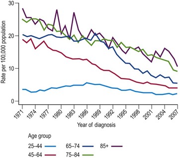
The incidence of cervical cancer in most countries has decreased significantly since the 1960s. In the UK, mortality from cervical cancer has been declining since 1950. The difference between mortality rates in 1950–1952 compared with 2005–2007 varies with age, from an extraordinary 85% reduction in women aged 55–64 years to a more moderate 15% reduction in women aged 25–34 years. Figure 34.1 shows the age-specific mortality rates for cervical cancer in the UK since 1971. It is seen that the greatest decreases have been in older women (aged ≥65 years), and that the increasing rates in younger women between 1970 and 1985 reversed in the 1990s.
The level of environmental exposure will be determined by social norms and will vary between ethnic groups and over time. Modelling shows that the idea that incidence and mortality rates can be modelled by age and cohort effects works well until the 1980s. However, more recent data require the addition of age-specific time trends corresponding to a beneficial effect of screening, particularly in younger women, to provide a satisfactory model (Sasieni and Adams 2000). From a public health perspective, it is important to note that women born in the 1960s are at three- to four-fold higher risk of cervical cancer compared with women born in the 1930s.
Risk factors
Evidence for an association between cervical cancer and sexual activity dates back to 1842, when Rigorni-Stern published data showing that whereas married women were more likely to die of cancer of the uterus (predominantly cervix) than breast cancer, nuns very rarely died of cancer of the uterus. Since then, the epidemiological evidence suggestive of a sexually transmitted agent causing cervical cancer has grown steadily. Traditional risk factors include the number of sexual partners and age at first sexual intercourse. The behaviour of men is also important, as shown by increasing risk in women with just one partner according to the number of partners of their husband (Buckley et al 1981). More recently, the sexually transmitted agent has been identified as certain types of HPV. The evidence that the relationship between HPV infection and cervical cancer is causal is overwhelming (Bosch et al 2002). Several large studies have been carried out to determine the prevalence of HPV in cervical cancer and precancerous lesions. Over 90% of cervical cancers have been found to include HPV DNA (Smith et al 2007), and when full adjustment for tissue adequacy and a range of polymerase chain reaction primers are used, the estimates rise to almost 100% (Walboomers et al 1999). Several studies have shown that HPV-negative women have an extremely low risk of CIN 3 or cancer (Bulkmans et al 2007, Cuzick et al 2008a, Dillner et al 2008). Furthermore, a recent study (Sankaranarayanan et al 2009) found no cancer deaths among 30,000 HPV-negative women in the subsequent 8-year period.
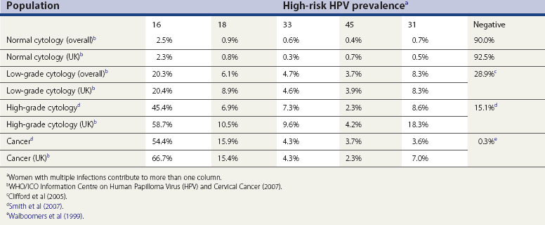
There are over 100 types of HPV, and only some of these infect the anogenital region. These can be split into low-risk types, which cause genital warts, and high-risk types, which can lead to cervical cancer. Types 16 and 18 are strongly associated with cervical cancer; other types including 31, 33, 35, 39, 45, 51, 52, 56, 58 and 66 are associated with a more moderate elevated risk. Table 34.2 details the prevalence of the most common high-risk types of HPV in women with normal cytology, precancerous cervical lesions and invasive cervical cancer in the UK and worldwide.
Table 34.2 High-risk human papilloma virus (HPV) prevalence in women with normal cytology, precancerous cervical lesions and invasive cervical cancer

Other risk factors are less clear cut. Smoking is generally found to be associated with cervical cancer, but it is difficult to disentangle the confounding caused by the sociological link between smoking and increased number of sexual partners. Nevertheless, the body of evidence available suggests that smoking increases the risk of cervical cancer two- to three-fold by reducing the local immune response to HPV (Kapeu et al 2009). The association between oral contraceptive use and cervical cancer is also confounded by sexual behaviour; however, a large pooled analysis including 24 studies found that women who had used oral contraceptives for 5 years or more were at almost double the risk of cervical cancer compared with women who had never used oral contraceptives. This risk declines after the use of oral contraceptives is stopped; within 10 years, the risk is similar to non-users (Appleby et al 2007). High parity and young age at first full-term pregnancy have, independently, been found to increase the risk of invasive cervical carcinoma, and this association remains after adjusting for other reproductive factors. Women with seven or more pregnancies are at approximately 78% greater risk than women with one or two pregnancies. It is estimated that cervical cancer could be reduced by 30% in developing countries if parity and age at first intercourse were the same as in developed countries (International Collaboration of Epidemiological Studies of Cervical Cancer 2006). Previous exposure to sexually transmitted diseases, in particular Chlamydia trachomatis and herpes simplex virus type 2, also increases the risk of cervical cancer, even after adjusting for HPV infection (Bosch and de Sanjose 2007).
Immunosuppression certainly conveys an increased risk of cervical cancer, as shown in studies on renal transplant patients receiving immunosuppressive drugs (Birkeland et al 1995) and on women who are human immunodeficiency virus (HIV) positive (Grulich et al 2007). It seems likely that diet plays a role in the immune response to HPV, but studies on diet and cervical cancer have found little evidence for a strong effect of intake of fruit and vegetables on the risk of cervical cancer (IARC Handbooks of Cancer Prevention 2003). It has been shown recently that cervical neoplasia (including CIS) exhibits familial clustering and that the strength of association increases with increasing genetic relatedness (Magnusson et al 1999). Independently, several groups have found an association between certain human leukocyte antigen class II antigens and cervical neoplasia. However, much more remains to be done in understanding the factors that determine why some women infected with oncogenic HPVs develop cervical cancer but the vast majority do not.
Natural history
There are few data from studies that directly observe the natural history of cervical cancer development because it is generally felt to be unethical not to treat precancerous cervical disease. The situation is further complicated by the possibility that the process of taking a biopsy, required for definitive diagnosis of disease, may affect the natural history by stimulating regression. Therefore, most of what is known of the natural history of cervical precancer is derived from the follow-up of women with cytological abnormalities and the study of the incidence and prevalence of cervical lesions. There is one exception — cervical CIS in Auckland, New Zealand between 1965 and 1974. Women with CIS were not treated. A judicial inquiry in 1988 concluded that this practice was unethical, but allowed the histological and other material to be used for further research. Two studies have been published using this data (McIndoe et al 1984, McCredie et al 2008), with the most recent paper including over 25 years of follow-up (McCredie et al 2008). This study provides the most direct estimates available of the rate of progression from CIS to invasive cancer; the results show that approximately 30% of CIS progress to cancer within 30 years.
For the vast majority (estimated as well over 95%) (Walboomers et al 1999) of cervical cancers, the first step is exposure to one of the oncogenic HPVs. The time from infection to the development of invasive cancer is thought to be many years; typically between 10 and 40. Longitudinal studies on young women show that the majority of HPV infections are transient (Moscicki et al 2004) and that the virus is indeed sexually transmitted (Burk et al 1996). Persistence of infection has been shown to be associated with the development of cervical lesions (Ho et al 1995). It is generally believed that one of the key steps in the development of cancer is integration of the viral DNA in the host genome (Das et al 1992), although some carcinomas only have episomal viral DNA (Cullen et al 1991).
Follow-up studies of women with CIN have found that approximately 60% of CIN I regresses compared with approximately 33% of CIN III; 11% and 22% of CIN I and II, respectively, progressed to CIN III (Östör 1993). Modellers find that regression is more common in younger women, and that three-quarters of CIN in women under 35 years of age will regress (van Ballegooijen et al 1997). They estimate the mean duration of CIN to be 12 years, and that the time from HPV infection to CIN is between 1 and 10 years. Although the details of progression and regression are largely speculative, it is clear that, at most, approximately one-third of high-grade CIN will progress to cancer over approximately 15 years and that the majority of CIN I will regress.
CIN III rates rise rapidly before 30 years of age. Rates then decrease, rather more slowly, being at approximately half their peak by 40 years of age and just 10–20% of their peak by 50 years of age (Office for National Statistics 2006). The extent to which published CIN III rates reflect the prevalence of an untreated condition and the extent to which they mirror incidence is not completely clear.
Prevention and screening
Estimates of the sensitivity of cytology are available from studies in which a large number of women with negative cytology have colposcopy. Ideally, colposcopy should be offered to all women, but this would be expensive and taking a biopsy from all women would be unethical. Many studies comparing two or more screening tests (such as cytology, HPV testing, direct visual inspection) offer colposcopy to all women who are positive on one or more of the screening tests. From such studies, the sensitivity of cytology for high-grade CIN is found to be between 50% and 75% (Nanda et al 2000, Cuzick et al 2006). However, most cases of missed CIN 3 would not become cancerous within 5 years. It is for this reason that screening is recommended, in most countries, to start around the age of 20 years and continue at regular intervals (of between 3 and 5 years) up to the age of 65 years.
There is now sufficient evidence that testing for HPV infections with a primary screening tool can reduce cervical cancer incidence and mortality rates (IARC Monographs 2007). Although HPV testing has high sensitivity (>90%), it is, on average, 6% less specific than cytology (Cuzick et al 2008b). This leads to a higher number of women being referred to colposcopy with no visible lesions. This is especially important in women under 30 years of age, in whom transient HPV infection and cervical lesions are most common. In many high-resource settings, HPV testing is being used in the triage of women over 30 years of age with borderline or mild smears. However, in low-resource settings where organized cytology is not feasible, introduction of HPV testing with subsequent ablative treatment for those women that are positive is an attractive alternative (Sankaranarayanan et al 2009). The introduction of HPV testing to the screening programme would allow the screening interval to be extended (Bulkmans et al 2007, Cuzick et al 2008b, Dillner et al 2008).
Human papilloma virus vaccines
Prophylactic HPV vaccines are L1 virus-like particles (VLPs) in which the surface L1 protein is made to aggregate into particles, mimicking the virus in antigenic respects but not containing the viral DNA. VLPs have been shown to induce a high titre of in-vitro neutralizing antibodies and to protect against experimental challenge with homologous virus in animal models. As VLPs do not contain any viral genes, they are non-infectious and non-oncogenic. To date, two vaccines are commercially available (Cervarix™ and Gardasil™); both are licensed to be applied in three doses within 6 months. Both preparations immunize against HPV 16 and 18, and Gardasil also immunizes against HPV 6 and 11. Results from a number of randomized controlled trials have been published to date. The phase III trials are designed to evaluate the endpoint of CIN 2/3 associated with the HPV types included in the vaccine (Harper et al 2006, Garland et al 2007, Paavonen et al 2007). The smaller phase II trials were powered primarily to evaluate protection from infection by the vaccine types (Villa et al 2005, Mao et al 2006).
There is no evidence that vaccination is effective against an HPV infection that is already present at the time of vaccination (i.e. vaccines do not induce HPV clearance or reduce progression) (Hildesheim et al 2007
Stay updated, free articles. Join our Telegram channel

Full access? Get Clinical Tree


