Abbott
Kimberly Clark
CORPAK (Viasys)
AMT
Bard
Percutaneous endoscopic gastrostomy (PEG) tubes
Flex-Flo® Introducer Gastrostomy Kit with Brown/Mueller T-Fastener™ Set 18 FR
MIC* percutaneous endoscopic gastrostomy feeding tube 14 FR
Corflo Max-PEG system 12, 16 and 20 FR
AMT Initial Placement Gastrsotmy Kit for use with 12–18 FR Low-Profile Gastrostomy Tube
Bard® PEG Feeding Devices
MIC* Safety PEG Feeding Tube/Kit, 14 FR O.D., push (over-the-wire) method
Balloon Replacement Gastrostomy Tubes
Easy-Feed® Gastrostomy Tubes 16–22 FR
MIC* Gastrostomy Feeding Tube 12–22 FR
CORFLO® Dual and Triple Gastrostomy Tubes
Bard® Tri-Funnel Replacement Gastrostomy Tube 12–24 FR
Magna-Port® Gastrostomy Tubes 14–26 FR
Dual—5 mL balloons—12, 14, 16, and 18 FR
20 mL balloons—16, 18, 20, 22 and 24 FR
Triple 20 mL balloons 16–24 FR
Balloon Low Profile Tubes
MIC-KEY* low-profile feeding tubes 12 FR 0.8 cm shaft to 24 FR 3.0 cm shaft
CORFLO-cuBBY low-profile gastrostomy 12, 14, 16, 18, 20 and 24 FR
AMT Mini ONE Button 12–24 FR, 0.8–6.5 cm shaft lengths
Dual Port WIZARD® Low-Profile Gastrostomy Device 16, 18, 20 and 24 FR with 10 AND 20 mL balloons
Diameter 12, 14, 16, 18, 20, 22 and 24 FR
Shaft lengths—1.0, 1.5, 2.0, 2.5, 3.0, 3.5, 4.0 and 4.5 cm, except the 12 FR tubes
16 FR has the maximum
Shaft length of 5.0 cm
Mushroom Low Profile Tube (button)
Capsule Non-Balloon Mini-ONE 14–24 FR
Bard® Button Replacement Gastrostomy Devices 18, 24, 28 FR
Obturated Non-Balloon Mini-ONE 14–24 FR
Gastro-Jejunal Tubes
MIC* Transgastric-Jejunal Feeding Tube-Endoscopic/Radiology Placement Kit, 16 FR
CORFLO®-Ultra Jejunostomy Tube
8 and 10 FR sizes for use with 20 FR PEGs
6 FR for use with 16 FR PEGs
G-JET Low Profile Gastric-Jejunal Enteral Tube
14, 16, 18 and 22 FR
MIC-KEY* Low-Profile Transgastric-Jejunal Feeding Tube 16 FR
MIC* Gastro-Enteric Feeding Tube, 16, 18, 20, 22 FR
Non-Balloon Replacement Gastrostomy Tubes
AMT Capsule Monarch G-Tube 12–20 FR
Ponsky™ Non-Balloon Replacement Gastrostomy Tubes 16, 20 FR
AMT Capsule Dome G-Tube 20FR
Jejunal Feeding Tubes
MIC-KEY* Low-Profile Jejunal Feeding Tube, 14 FR O.D., 0.8 cm stoma length, 5 mL balloon
MIC Jejunal Feeding Tube 12–24 FR
Care of the gastrostomy post placement is simple. The skin around the G-tube should be washed daily with a mild soap and water. Use of hydrogen peroxide should be avoided as it causes unnecessary irritation to the skin. If erythema is present, using a topical antibiotic ointment may be indicated. Covering the skin with gauze should be avoided.
Complications of Gastrostomy/Gastrojejunostomy Tubes
The most commonly encountered complications of enteral devices include infection, leakage, granulation tissue, migration, obstruction, and persistent fistula after removal [3].
Infection
Gastrostomy tube infections are more common in the first several weeks following percutaneous placement. It has been estimated that 25–33 of patients develop a peristomal infection [4]. Few studies have addressed the issue of peristomal infections in children. The underlying medical condition of the child may influence their risk for infection and hinder wound healing. Antibiotic prophylaxis with placement of percutaneous endoscopic gastrostomies is recommended by the American Society for Gastrointestinal Endoscopy [5, 6].
Infection of the peristomal area can present with a variety of symptoms. Fever, spreading erythema, tenderness, pain, induration, and purulent discharge are typical. However, the yellow–brown crusty discharge that is commonly seen around the gastrostomy site is not a sign of infection, a finding that is confusing to families and caregivers. In case of infection treatment with a topical antibiotic may be all that is needed, however oral antibiotics may be necessary. Most infections respond to a first generation cephalosporin.
Abscess formation adjacent to the stoma is another potential complication. These lesions have a rapid onset of a pustule or a red–purple fluid-filled lesion that is tender to the touch. When it ruptures, a punctuate opening is apparent and may drain for several days. Treatment with warm compresses and antibiotic therapy is recommended.
Feeding tubes may become colonized with microbial organisms, yeast, and fungus. There have been more 100 different microorganisms isolated from gastrostomy tubes with the most common being Candida (Fig. 40.1), Pseudomonas, Escherichia coli, Enterobacter cloacae, Streptococci, Lactobacillus, Staphylococcus aureus, and Bacteroides. The significance of gastrostomy tube colonization is unclear; however in the face of recurrent infections, culture of the site and treatment with the appropriately sensitive antibiotic is recommended.


Fig. 40.1
Fungal tube site infection
Tube Migration
Migration of the gastrostomy tube with aberrant tract formation has been reported. The buried bumper syndrome (retrograde migration of the gastrostomy tube’s internal bumper into the abdominal wall or into the stoma tract) is well described [7]. This occurs when there is traction placed on the external portion of the gastrostomy tube that results in excessive tension on the internal bumper at the time of placement. A false tract may develop as a late complication when the shaft length of the low-profile gastrostomy tube is not resized in a growing child. Failure to remeasure the shaft length may result in a too short tube causing the balloon or internal bumper to move up into the tract. Leakage and focal abdominal discomfort may result. Long-term migration of the balloon in to the tract may result in the development of a false tract or dilatation of the gastric opening. This allows for drainage of gastric contents onto the skin resulting in peristomal skin excoriation and breakdown.
It is important to remeasure the stromal tract correctly and accurately. It is best to measure with the patient in both a supine and sitting position. If the measure significantly differs in either position, the tract length should be the average between the two measurements. It is recommended that tracts be remeasured annually.
Leakage
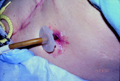
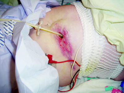
Chronic leakage is a worrisome complication (Fig. 40.2). Leakage of gastric contents will result in peristomal skin breakdown and the subsequent pain and potential infection. The first goal is always to stop the leakage. Remeasuring the shaft length and ensuring the proper fit of the gastrostomy tube is the first step. Ensuring that there is adequate water in the balloon is important. While most manufacturers recommend that the balloon be inflated with 4–6 mL of water, most can accommodate up to 10 mL safely. It is important to be cognizant of the size of the child and their gastric volume, to avoid exceeding gastric capacity. If the leakage is from an enlarged stoma tract, increasing the diameter of the gastrostomy tube (for example, going from a 14 to a 16 French) may be necessary. However, continuing to increase the size of the tube should be avoided when possible. Removing the gastrostomy tube for a couple of hours and allowing the stoma to “shrink” may be helpful. Many patients benefit from changing to a different appliance if leakage is a chronic issue. If all interventions fail, surgical revision may be necessary.


