Chapter 25 Endometriosis and Adenomyosis
Endometriosis and adenomyosis often present difficult diagnostic challenges. In the case of endometriosis, few gynecologic conditions require such difficult surgical dissections.
Endometriosis
Endometriosis is a benign condition in which endometrial glands and stroma are present outside the uterine cavity and walls. Endometriosis is important in gynecology because of its frequency, distressing symptomatology, association with infertility, and potential for invasion of adjacent organ systems, such as the gastrointestinal or urinary tracts.
OCCURRENCE
The prevalence of endometriosis in the general population is not known, but it is estimated that 5% to 15% of women have some degree of the disease. At least one third of women with chronic pelvic pain have visualized endometriosis, as do a significant number of infertile women. Interestingly, endometriosis is noted in 5% to 15% of women undergoing gynecologic laparotomies, and it is an unexpected finding in about half of these cases.
The typical patient with endometriosis is in her 30s, nulliparous, and infertile. However, in practice, many women with endometriosis do not fit the classic picture. Occasionally, endometriosis may occur in infancy, childhood, or adolescence, but at these early ages, it is usually associated with obstructive genital anomalies such as a uterine or vaginal septum. Although endometriosis should regress after menopause unless estrogens are prescribed, 5% of new cases develop in that age group. In addition, the scarifying involution from preexisting lesions may result in obstructive problems, especially in the gastrointestinal and urinary tracts.
PATHOGENESIS
The pathogenesis of endometriosis is not completely understood. Genetic predisposition clearly plays a role. The following three hypotheses have been used to explain the various manifestations of endometriosis and the different locations in which endometriotic implants may be found:
Most authorities today believe that several factors are involved in the initiation and spread of endometriosis, including retrograde menstruation, coelomic metaplasia, immunologic changes, and genetic predisposition. A fundamental question is why all menstruating women do not develop endometriosis given that most if not all women have retrograde flow into the pelvis during menstruation. The amount of exposure to retrograde flow and the woman’s immunologic response appear to be critical. Researchers have identified differences in the chemical composition and biologic pathways of endometrial cells from women with endometriosis compared with unaffected women. They have also found significant differences in the inflammatory factors and growth factors in the peritoneal fluid of affected women. A clearer understanding of the pathophysiology of endometriosis would provide insights into more effective strategies for prevention and treatment.
SITES OF OCCURRENCE
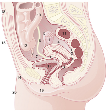
Endometriosis occurs most commonly in the dependent portions of the pelvis. Specifically, implants can be found on the ovaries, the broad ligament, the peritoneal surfaces of the cul-de-sac (including the uterosacral ligaments and posterior cervix), and the rectovaginal septum (Figure 25-1). Quite frequently, the rectosigmoid colon is involved, as is the appendix and the vesicouterine fold of peritoneum. Endometriosis is occasionally seen in laparotomy scars, developing especially after a cesarean delivery or myomectomy when the endometrial cavity has been entered. It is probable that endometrial tissue is seeded into the surgical incision. Two of three women with endometriosis have ovarian involvement.

FIGURE 25-1 Common sites of endometriosis in decreasing order of frequency: (1) ovary, (2) cul-de-sac, (3) uterosacral ligaments, (4) broad ligaments, (5) fallopian tubes, (6) uterovesical fold, (7) round ligaments, (8) vermiform appendix, (9) vagina, (10) rectovaginal septum, (11) rectosigmoid colon, (12) cecum, (13) ileum, (14) inguinal canals, (15) abdominal scars, (16) ureters, (17) urinary bladder, (18) umbilicus, (19) vulva, and (20) peripheral sites.
PATHOLOGY
Islands of endometriosis respond cyclically to ovarian steroidal hormone production. The implants proliferate under estrogenic stimulation and slough when support from estrogen and progesterone is removed with involution of the corpus luteum. The sloughed material induces a profound inflammatory response resulting immediately in pain and fibrosis in the long term. The macroscopic appearance of endometriosis depends on the site of the implant, activity of the lesion, day of the menstrual cycle, and time since implantation.
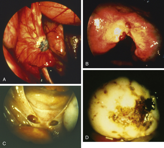
Lesions may be raised and flat with red, black, or brown coloration; fibrotic scarred areas that are yellow or white in hue; or vesicles that are pink, clear, or red (Figure 25-2). The color of the implant is generally determined by its vascularity, the size of the lesion, and the amount of residual sloughed material. Newer implants tend to be red, blood-filled active lesions. Older lesions tend to be much less active hormonally, scarred and blue-gray in color with a puckered appearance. These older inactive lesions have been called the “tattooing of endometriosis.”

FIGURE 25-2 A to D: Appearance of old endometriosis with “tattooing” (blue-gray lesions), and red, brown, and black raised lesions of active endometriosis at the time of laparoscopy.
Endometriomas of the ovary are cysts filled with thick, chocolate-colored fluid that sometimes has the black color and tarry consistency of crankcase oil. This characteristic fluid represents aged, hemolyzed blood and desquamated endometrium. Usually, endometrial glands and stroma are present in the cyst wall. Sometimes, however, the pressure of the enclosed fluid destroys the endometrial lining of the endometrioma, leaving only a fibrotic cyst wall infiltrated with large numbers of hemosiderin-laden macrophages. Generally, ovarian implants are associated with significant scarring of the ovary to the pelvic side wall or broad ligament. Histologically, two of four characteristics must be found in the endometrioma specimen to confirm the diagnosis—endometrial epithelium, endometrial glands, endometrial stroma, and hemosiderin-laden macrophages.
Although endometriosis is a benign process, it shares many characteristics with malignancy. It is locally infiltrative, invasive, and widely disseminated. It is also curious that cyclic hormones tend to induce growth, whereas continuous hormonal exposure, especially in high doses, generally induces significant regression.
STAGING
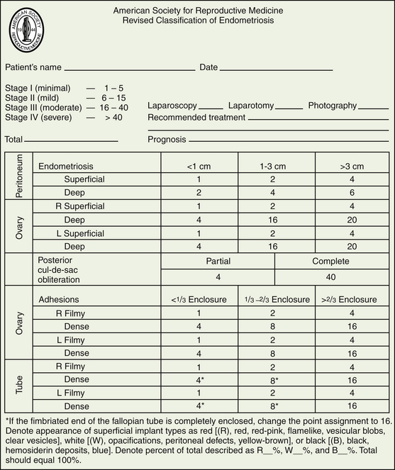
The American Society of Reproductive Medicine employs a staging protocol in an attempt to correlate fertility potential with a quantified stage of endometriosis. This staging, which was initially based on the allocation of points depending on the sites involved and extent of visualized disease (Figure 25-3), was modified to include a description of the color of the lesions and the percentage of surface involved in each lesion type, as well as a more detailed description of any endometrioma.

Stay updated, free articles. Join our Telegram channel

Full access? Get Clinical Tree

