Chapter 5 Endocrinology of Pregnancy and Parturition
Women undergo major endocrinologic and metabolic changes that establish, maintain, and terminate pregnancy. The aim of these changes is the safe delivery of an infant who can survive outside of the uterus. The maturation of the fetus and the adaptation of the mother are regulated by a variety of hormones. This chapter deals with the properties, functions, and interactions of the most important of these hormones as they relate to pregnancy and parturition.
Fetoplacental Unit
The concept of the fetoplacental unit is based on observations of the interactions of hormones of fetal and maternal origin. The fetoplacental unit largely controls the endocrine events of the pregnancy. Although the fetus, the placenta, and the mother all provide input, the fetus appears to play the most active and controlling role of the three in its growth and maturation, and probably also in the events that lead to parturition.
FETUS
The adrenal gland is the major endocrine component of the fetus. In mid-pregnancy, it is larger than the fetal kidney. The fetal adrenal cortex consists of an outer definitive, or adult, zone and an inner, fetal, zone. The definitive zone later develops into the three components of the adult adrenal cortex: the zona fasciculata, the zona glomerulosa, and the zona reticularis. During fetal life, the definitive zone secretes primarily glucocorticoids and mineralocorticoids. The fetal zone, at term, constitutes 80% of the fetal gland and primarily secretes androgens during fetal life. It involutes following delivery and completely disappears by the end of the first year of life. The fetal adrenal medulla synthesizes and stores catecholamines, which play an important role in maintaining fetal homeostasis. The role of the fetal adrenal during fetal growth and maturation is not completely understood.
PLACENTA
The placenta produces both steroid and peptide hormones in amounts that vary with gestational age. Precursors for progesterone synthesis come from the maternal circulation. Because of the lack of the enzyme 17α-hydroxylase, the human placenta cannot directly convert progesterone to estrogen but must use androgens, largely from the fetal adrenal gland, as its source of precursor for estrogen production.
MOTHER
The mother adapts to pregnancy through major endocrinologic and metabolic changes. The ovaries produce progesterone in early pregnancy until its production shifts to the placenta. The maternal hypothalamus and posterior pituitary produce and release oxytocin, which causes uterine contractions and milk letdown. The anterior pituitary produces prolactin, which stimulates milk production. Several important changes in maternal metabolism are described later in the chapter.
Hormones
The fetoplacental unit produces a variety of hormones to support the maturation of the fetus and the adaptation of the mother.
PEPTIDE HORMONES
Human Chorionic Gonadotropin
Human chorionic gonadotropin (hCG) is secreted by trophoblastic cells of the placenta and maintains pregnancy. This hormone is a glycoprotein with a molecular weight of 40,000 to 45,000 and consists of two subunits: alpha (α) and beta (β). The α subunit is shared with luteinizing hormone (LH) and thyroid-stimulating hormone (TSH). The specificity of hCG is related to its β subunit (β-hCG), and a radioimmunoassay that is specific for the β subunit allows positive identification of hCG. The presence of hCG at times other than pregnancy signals the presence of an hCG-producing tumor, usually a hydatidiform mole, choriocarcinoma, or embryonal carcinoma (a germ cell tumor).
During pregnancy, hCG begins to rise 8 days after ovulation (9 days after the midcycle LH peak). This provides the basis for virtually all immunologic or chemical pregnancy tests. With continuing pregnancy, hCG values peak at 60 to 90 days and then decline to a moderate, more constant level. For the first 6 to 8 weeks of pregnancy, hCG maintains the corpus luteum and thereby ensures continued progesterone output until progesterone production shifts to the placenta. Titers of hCG are usually abnormally low in patients with an ectopic pregnancy or threatened abortion and abnormally high in those with trophoblastic disease (e.g., moles or choriocarcinoma). This hormone may also regulate steroid biosynthesis in the placenta and the fetal adrenal gland and stimulate testosterone production in the fetal testicle. Although immune suppression has been ascribed to hCG, this effect has not been verified.
Human Placental Lactogen
Human placental lactogen (hPL) originates in the placenta. It is a single-chain polypeptide with a molecular weight of 22,300, and it resembles pituitary growth hormone and human prolactin in structure. Maternal serum concentrations parallel placental weight, rising throughout gestation to maximum levels in the last 4 weeks. At term, hPL accounts for 10% of all placental protein production. Low values are found with threatened abortion and intrauterine fetal growth restriction. Human placental lactogen antagonizes the cellular action of insulin and decreases maternal glucose utilization, which increases glucose availability to the fetus. This may play a role in the pathogenesis of gestational diabetes.
Corticotropin-Releasing Hormone
During pregnancy the major source of corticotropin-releasing hormone (CRH) is the placenta, and it can be measured as early as 12 weeks of gestation when it passes into the fetal circulation. This 41–amino acid peptide stimulates fetal adrenocorticotropic hormone (ACTH) secretion, which in turn stimulates the fetal adrenal to secrete dehydroepiandrosterone sulfate (DHEA-S), an important precursor of estrogen production by the placenta. The fetal adrenal gland early in pregnancy does not have the enzymes to produce cortisol, but as gestational age increases, it becomes more responsive. Fetal cortisol stimulates placental CRH release, which then stimulates fetal ACTH secretion, completing a positive feedback loop that plays an important role in the activation and amplification of labor, both preterm and term. Elevated levels of CRH in mid-gestation have been found to be associated with an increased risk for subsequent preterm labor.
Prolactin
Prolactin is a peptide from the anterior pituitary with a molecular weight of about 20,000. Normal nonpregnant levels are about 10 ng/mL. During pregnancy, maternal prolactin levels rise in response to increasing maternal estrogen output that stimulates the anterior pituitary lactotrophs. The main effect of prolactin is stimulation of postpartum milk production. In the second half of pregnancy, prolactin secreted by the fetal pituitary may be an important stimulus of fetal adrenal growth. Prolactin may also play a role in fluid and electrolyte shifts across the fetal membranes.
STEROID HORMONES
Progesterone
Progesterone is the most important human progestogen. In the luteal phase, it induces secretory changes in the endometrium, and in pregnancy, higher levels induce decidual changes. Up to the 6th or 7th week of pregnancy, the major source of progesterone (in the form of 17-OH progesterone) is the ovary. Thereafter, the placenta begins to play the major role. If the corpus luteum of pregnancy is removed before 7 weeks and continuation of the pregnancy is desired, progesterone should be given to prevent spontaneous abortion. Circulating progesterone is mostly bound to carrier proteins, and less than 10% is free and physiologically active.
The myometrium receives progesterone directly from the venous blood draining the placenta. Progesterone prevents uterine contractions and may also be involved in establishing an immune tolerance for the products of conception. Progesterone also suppresses gap junction formation, placental CRH expression, and the actions of estrogen, cytokines, and prostaglandin. This steroid hormone therefore plays a central role in maintaining uterine quiescence throughout most of pregnancy.
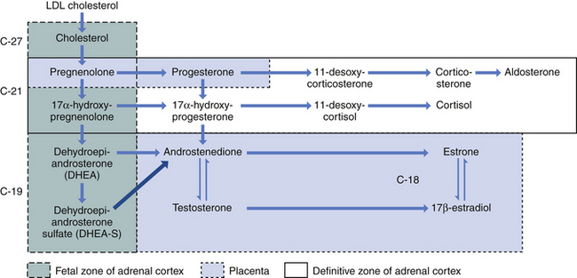
The fetus inactivates progesterone by transformation to corticosteroids or by hydroxylation or conjugation to inert excretory products. However, the placenta can convert these inert materials back to progesterone. Steroid biochemical pathways are shown in Figure 5-1.
Estrogens
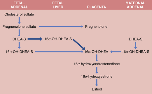
Both fetus and placenta are involved in the biosynthesis of estrone, estradiol, and estriol. Cholesterol is converted to pregnenolone and pregnenolone sulfate in the placenta. These precursors are converted to DHEA-S largely in the fetal, and to a lesser extent the maternal, adrenals. The DHEA-S is further metabolized by the placenta to estrone (E1) and, through testosterone, to estradiol (E2). Estriol (E3), the most abundant estrogen in human pregnancy, is synthesized in the placenta from 16α-hydroxy-DHEA-S, which is produced in the fetal liver from adrenal DHEA-S. Placental sulfatase is required to deconjugate 16α-hydroxy-DHEA-S before conversion to E3 (Figure 5-2). Steroid sulfatase activity in the placenta is high except in rare cases of sulfatase deficiency.
A sudden decline of estriol in the maternal circulation may indicate fetal compromise in neurologically intact fetuses. Anencephalic fetuses lack a hypothalamus and have hypoplastic anterior pituitary and adrenal glands; thus, estriol production is only about 10% of normal.

Full access? Get Clinical Tree

