Electrolyte and Acid-Base Disorders

Chapter 52 Electrolyte and Acid-Base Disorders


52.1 Composition of Body Fluids


Total Body Water


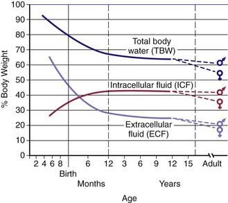
Water is the most plentiful constituent of the human body. Total body water (TBW) as a percentage of body weight varies with age (Web imageFig. 52-1). The fetus has very high TBW, which gradually decreases to approximately 75% of birthweight for a term infant. Premature infants have higher TBW than term infants. During the 1st yr of life, TBW decreases to approximately 60% of body weight and basically remains at this level until puberty. At puberty, the fat content of females increases more than that of males, who acquire more muscle mass than females. Because fat has very low water content and muscle has high water content, by the end of puberty TBW in males remains at 60%, but TBW in females decreases to approximately 50% of body weight. The high fat content in overweight children causes a decrease in TBW as a percentage of body weight. During dehydration, TBW decreases and, thus, is a smaller percentage of body weight.


Fluid Compartments


TBW is divided between 2 main compartments: intracellular fluid (ICF) and extracellular fluid (ECF). In the fetus and newborn, the ECF volume is larger than the ICF volume (see Web Fig. 52-1). The normal postnatal diuresis causes an immediate decrease in the ECF volume. This is followed by continued expansion of the ICF volume, which results from cellular growth. By 1 yr of age, the ratio of the ICF volume to the ECF volume approaches adult levels. The ECF volume is approximately 20-25% of body weight, and the ICF volume is approximately 30-40% of body weight, close to twice the ECF volume (Web Fig. 52-2). With puberty, the increased muscle mass of males causes them to have a higher ICF volume than females. There is no significant difference in the ECF volume between postpubertal females and males.
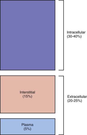


The ECF is further divided into the plasma water and the interstitial fluid (see Web Fig. 52-2). The plasma water is 5% of body weight. The blood volume, given a hematocrit of 40%, is usually 8% of body weight, although it is higher in newborns and young infants; in premature newborns, it is approximately 10% of body weight. The volume of plasma water can be altered by pathologic conditions, including dehydration, anemia, polycythemia, heart failure, abnormal plasma osmolality, and hypoalbuminemia. The interstitial fluid, normally 15% of body weight, can increase dramatically in diseases associated with edema, such as heart failure, protein-losing enteropathy, liver failure, nephrotic syndrome, and sepsis. An increase in interstitial fluid also occurs in patients with ascites or pleural effusions.


There is normally a delicate equilibrium between the intravascular fluid and the interstitial fluid. The balance between hydrostatic and oncotic forces regulates the intravascular volume, which is critical for proper tissue perfusion. The intravascular fluid has a higher concentration of albumin than the interstitial fluid, and the consequent oncotic force draws water into the intravascular space. The maintenance of this gradient depends on the limited permeability of albumin across the capillaries. The hydrostatic pressure of the intravascular space, which is due to the pumping action of the heart, drives fluid out of the intravascular space. These forces favor movement into the interstitial space at the arterial ends of the capillaries. The decreased hydrostatic forces and increased oncotic forces, which result from the dilutional increase in albumin concentration, cause movement of fluid into the venous ends of the capillaries. Overall, there is usually a net movement of fluid out of the intravascular space to the intracellular space, but this fluid is returned to the circulation via the lymphatics. An imbalance in these forces may cause expansion of the interstitial volume at the expense of the intravascular volume. In children with hypoalbuminemia, the decreased oncotic pressure of the intravascular fluid contributes to the development of edema. Loss of fluid from the intravascular space may compromise the intravascular volume, placing the child at risk for inadequate blood flow to vital organs. This is especially likely in diseases in which capillary leak occurs because the loss of albumin from the intravascular space is associated with an increase in the albumin concentration in the interstitial space, further compromising the oncotic forces that normally maintain intravascular volume. In contrast, with heart failure, there is an increase in venous hydrostatic pressure from expansion of the intravascular volume, which is caused by impaired pumping by the heart, and the increase in venous pressure causes fluid to move from the intravascular space to the interstitial space. Expansion of the intravascular volume and increased intravascular pressure also cause the edema that occurs with acute glomerulonephritis.


Electrolyte Composition


The composition of the solutes in the ICF and ECF are very different (Web Fig. 52-3). Sodium and chloride are the dominant cation and anion, respectively, in the ECF. The sodium and chloride concentrations in the ICF are much lower. Potassium is the most abundant cation in the ICF, and its concentration within the cells is approximately 30 times higher than in the ECF. Proteins, organic anions, and phosphate are the most plentiful anions in the ICF. The dissimilarity between the anions in the ICF and the ECF is largely determined by the presence of intracellular molecules that do not cross the cell membrane, the barrier separating the ECF and the ICF. In contrast, the difference in the distribution of cations—sodium and potassium—is due to the activity of the Na+,K+-ATPase pump, which uses cellular energy to actively extrude sodium from cells and move potassium into cells. The chemical gradient between the intracellular potassium concentration and the extracellular potassium concentration creates the electrical gradient across the cell membrane. The concentration-dependent movement of potassium out of the cell makes the intracellular space negative relative to the extracellular space.
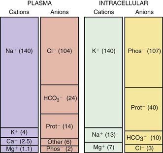


The difference in the electrolyte compositions of the ECF and the ICF has important ramifications in the evaluation and treatment of electrolyte disorders. The serum concentration of an electrolyte, which is measured clinically, does not always reflect the body content. This is due to the larger volume of the ICF compared with the ECF and the variation in electrolyte concentrations between these 2 compartments. The intracellular potassium concentration is much higher than the serum concentration. A shift of potassium from the intracellular space can maintain a normal or even an elevated serum potassium concentration, despite massive losses of potassium from the intracellular space. This is dramatically seen in diabetic ketoacidosis, in which a state of significant potassium depletion is often masked because of a transmembrane shift of potassium from the ICF to the ECF. For potassium and phosphorus, electrolytes with a high intracellular concentration, the serum level may not reflect total body content. Similarly, the serum calcium concentration does not predict the body content of calcium, which is largely in bone.


Osmolality


The ICF and the ECF are in osmotic equilibrium because the cell membrane is permeable to water. If the osmolality in 1 compartment changes, then water movement leads to a rapid equalization of osmolality. This can lead to significant shifts of water between the intracellular space and the extracellular space. Clinically, the primary process is usually a change in the osmolality of the ECF, with a resultant shift of water into the ICF if the ECF osmolality decreases or a shift of water out of the ICF if the ECF osmolality increases. The osmolality of the ECF can be determined, and it usually equals the ICF osmolality. The plasma osmolality is normally 285-295 mOsm/kg, and it is measured by the degree of freezing point depression. The plasma osmolality can also be estimated by a calculation based on the following formula:


image


Glucose and blood urea nitrogen (BUN) are measured in mg/dL. Division of these values by 18 and 2.8, respectively, as shown, converts the units into mmol/L. Multiplication of the sodium value by 2 accounts for its accompanying anions, principally chloride and bicarbonate. The calculated osmolality is usually slightly lower than the measured osmolality.


Glucose and urea normally contribute little to the plasma osmolality; multiplication of the sodium value by 2 provides an approximation of the osmolality. Urea is not confined to the extracellular space because it readily crosses the cell membrane and its intracellular concentration approximately equals its extracellular concentration. Whereas an elevated sodium concentration causes a shift of water from the intracellular space, with uremia, there is no osmolar gradient between the 2 compartments and, consequently, no movement of water. The only exception is during hemodialysis, when the decrease in extracellular urea is so rapid that the intracellular urea does not have time to equilibrate. This may lead to the disequilibrium syndrome, in which water shifts into brain cells, potentially causing severe symptoms. Ethanol, because it freely crosses cell membranes, is another ineffective osmole. The effective osmolality can be calculated as follows:


image


The effective osmolality (also called the tonicity) determines the osmotic force that is mediating the shift of water between the ECF and the ICF.


Hyperglycemia causes an increase in the plasma osmolality because it is not in equilibrium with the intracellular space. During hyperglycemia there is a shift of water from the intracellular space to the extracellular space. This is clinically important in children with hyperglycemia during diabetic ketoacidosis. The shift of water causes dilution of the sodium in the extracellular space, causing hyponatremia despite an elevated plasma osmolality. The magnitude of this effect can be calculated as follows:


image


where [Na]measured = sodium concentration measured by the clinical laboratory and [Na]corrected = corrected sodium concentration (the sodium concentration if the glucose concentration were normal and its accompanying water moved back into the cells). The [Na]corrected is the more reliable indicator of the patient’s true ratio of total body sodium to TBW, the normal determinant of the sodium concentration.


Normally, the measured osmolality and the calculated osmolality are within 10 mOsm/kg. However, there are some clinical situations in which this does not occur. The presence of unmeasured osmoles causes the measured osmolality to be significantly elevated in comparison with the calculated osmolality. This difference is the osmolal gap, which is present when the measured osmolality exceeds the calculated osmolality by >10 mOsm/kg. Examples of unmeasured osmoles include ethanol, ethylene glycol, methanol, and mannitol. These substances increase the measured osmolality but are not part of the equation for calculating osmolality. The presence of an osmolal gap is a clinical clue to the presence of unmeasured osmoles and may be diagnostically useful when there is clinical suspicion of poisoning with methanol or ethylene glycol.


Pseudohyponatremia is a second situation in which there is discordance between the measured osmolality and the calculated osmolality. Lipids and proteins are the solids of the serum. In patients with elevated serum lipids or proteins, the water content of the serum decreases because water is displaced by the larger amount of solids. Some clinical laboratories measure sodium concentration by determining the amount of sodium per liter of serum, including the solid component. When the solid component increases, there is a decrease in the sodium concentration per liter of serum, despite a normal concentration of sodium when based on the amount of sodium per liter of serum water. It is the concentration of sodium in serum water that is physiologically relevant. In such situations, the plasma osmolality is normal despite the presence of pseudohyponatremia, because the method for measuring osmolality is not appreciably influenced by the percentage of serum that is composed of lipids and proteins. Pseudohyponatremia is diagnosed by the finding of a normal measured plasma osmolality despite hyponatremia. This laboratory artifact does not occur if the sodium concentration in water is measured directly with an ion-specific electrode, such as occurs with the instruments used for measuring arterial blood gases.


When there are no unmeasured osmoles and pseudohyponatremia is not a concern, the calculated osmolality provides an accurate estimate of the plasma osmolality. Measurement of plasma osmolality is useful for detecting or monitoring unmeasured osmoles and confirming the presence of true hyponatremia. Whereas many children with high plasma osmolality are dehydrated—as seen with hypernatremic dehydration or diabetic ketoacidosis—high osmolality does not always equate with dehydration. A child with salt poisoning or uremia has an elevated plasma osmolality but may be volume overloaded. In many situations, it is best to focus on the components of the plasma osmolality and to analyze them individually to reach a correct clinical conclusion.


52.2 Regulation of Osmolality and Volume


The regulation of plasma osmolality and the intravascular volume are controlled by independent systems for water balance, which determines osmolality, and sodium balance, which determines volume status. Maintenance of normal osmolality depends on control of water balance. Control of volume status depends on regulation of sodium balance. When volume depletion is present, it takes precedence over regulation of osmolality, and retention of water contributes to the maintenance of intravascular volume.


Regulation of Osmolality


The plasma osmolality is tightly regulated and maintained at 285-295 mOsm/kg. Modification of water intake and excretion maintains normal plasma osmolality. In the steady state, the combination of water intake and water produced by the body from oxidation balances water losses from the skin, lungs, urine, and gastrointestinal tract. Only water intake and urinary losses can be regulated.


Osmoreceptors in the hypothalamus sense the plasma osmolality (Chapter 552). An elevated effective osmolality leads to secretion of antidiuretic hormone (ADH) by neurons in the supraoptic and paraventricular nuclei in the hypothalamus. The axons of these neurons terminate in the posterior pituitary. Circulating ADH binds to its V2 receptors in the collecting duct cells of the kidney, and, via the generation of cyclic adenosine monophosphate, causes insertion of water channels (aquaporin-2) into the renal collecting ducts. This produces increased permeability to water, permitting resorption of water into the hypertonic renal medulla. The end result is that the urine concentration increases and water excretion decreases. Urinary water losses cannot be completely eliminated because there is obligatory excretion of urinary solutes, such as urea and sodium. The regulation of ADH secretion is tightly linked to plasma osmolality, responses being detectable with a 1% change in the osmolality. ADH secretion virtually disappears when the plasma osmolality is low, allowing excretion of maximally dilute urine. The consequent loss of free water (water without sodium) corrects the plasma osmolality. ADH secretion is not an all-or-nothing response; there is a graded adjustment as the osmolality changes.


Production of concentrated urine under the control of ADH requires a hypertonic renal medulla. The countercurrent multiplier, produced by the loop of Henle and the vasa recta, generates this hypertonicity. ADH stimulates sodium transport in the loop of Henle, helping to maintain this gradient when water retention is necessary.


Water intake is regulated by hypothalamic osmoreceptors, although these are different from the osmoreceptors that determine ADH secretion. These hypothalamic osmoreceptors, by linking to the cerebral cortex, stimulate thirst when the serum osmolality increases. Thirst occurs with a small increase in the serum osmolality.


Control of osmolality is subordinate to maintenance of an adequate intravascular volume. When volume depletion is present, both ADH secretion and thirst are stimulated, regardless of the plasma osmolality. The sensation of thirst requires moderate volume depletion but only a 1-2% change in the plasma osmolality. Although all of the mechanisms are not clear, angiotensin II, which is increased during volume depletion, is known to stimulate thirst. Baroreceptors, when sensing volume depletion, may also stimulate thirst.


A number of conditions can limit the kidney’s ability to excrete adequate water to correct low plasma osmolality. In the syndrome of inappropriate antidiuretic hormone (SIADH), ADH continues to be produced despite a low plasma osmolality. In the presence of ADH, urinary dilution does not occur, and sufficient water is not excreted (Chapters 52.3 and 553).


The glomerular filtration rate (GFR) affects the kidney’s ability to eliminate water. With a decrease in the GFR, less water is delivered to the collecting duct, limiting the amount of water that can be excreted. The impairment in the GFR must be quite significant to limit the kidney’s ability to respond to an excess of water.


The minimum urine osmolality is approximately 30-50 mOsm/kg. This places an upper limit on the kidney’s ability to excrete water; sufficient solute must be present to permit water loss. Massive water intoxication may exceed this limit, whereas a lesser amount of water is necessary in the child with a diet that has very little solute. This is occasionally seen and can produce severe hyponatremia in children who receive little salt and have little urea production as a result of inadequate protein intake. Volume depletion is an extremely important cause of decreased water loss by the kidney despite a low plasma osmolality. This “appropriate” secretion of ADH occurs because volume depletion takes precedence over the osmolality in the regulation of ADH.


The normal response to increased plasma osmolality is conservation of water by the kidney. In central diabetes insipidus, this does not occur because of an absence of ADH secretion (Chapter 552.1). Patients with nephrogenic diabetes insipidus have an inability to respond to ADH and produce dilute urine despite an increase in plasma osmolality (Chapters 52.3, 524, and 552).


The maximum urine osmolality is about 1,200 mOsm/kg. The obligatory solute losses dictate the minimum volume of urine that must be produced, even when maximally concentrated. Obligatory water losses increase in patients with high salt intake or high urea losses, as may occur after relief of a urinary obstruction or during recovery from acute tubular necrosis. An increase in urinary solute and, consequently, water losses occurs with an osmotic diuresis, which occurs classically from glycosuria in diabetes mellitus as well as iatrogenically after mannitol administration. There are developmental changes in the kidney’s ability to concentrate the urine. The maximum urine osmolality in a newborn, especially a premature newborn, is less than that in an older infant or child. This limits the ability to conserve water and makes such a patient more vulnerable to hypernatremic dehydration. Very high fluid intake, as seen with psychogenic polydipsia, can dilute the high osmolality in the renal medulla, which is necessary for maximal urinary concentration. If fluid intake is restricted in patients with this condition, there may be some impairment in the kidney’s ability to concentrate the urine, although this defect corrects after a few days without polydipsia. This may also occur during the initial treatment of central diabetes insipidus with desmopressin acetate; the renal medulla takes time to achieve its normal maximum osmolality. Loop diuretics, such as furosemide, by inhibiting sodium resorption in the ascending limb of the loop of Henle, decrease medullary hypertonicity, preventing excretion of maximally concentrated urine.


Regulation of Volume


An appropriate intravascular volume is critical for survival; both volume depletion and volume overload may cause significant morbidity and mortality. Because sodium is the principal extracellular cation and it is restricted to the ECF, adequate body sodium is necessary for maintenance of intravascular volume. The principal extracellular anion, chloride, is also necessary, but for simplicity, sodium balance is considered the main regulator of volume status because body content of sodium and that of chloride usually change proportionally, given the need for equal numbers of cations and anions. In some situations, chloride depletion is considered the dominant derangement causing volume depletion (metabolic alkalosis with volume depletion). In other situations, such as volume depletion with metabolic acidosis, sodium depletion may exceed chloride depletion.


The kidney determines sodium balance because there is little homeostatic control of sodium intake, even though salt craving does occasionally occur, typically in children with chronic renal salt loss. The kidney regulates sodium balance by altering the percentage of filtered sodium that is resorbed along the nephron. Normally, the kidney excretes <1% of the sodium filtered at the glomerulus. In the absence of disease, extrarenal losses and urinary output match intake, with the kidney having the capacity to adapt to large variations in sodium intake. When necessary, urinary sodium excretion can be reduced to virtually undetectable levels or increased dramatically.


Urinary sodium excretion is regulated by both intrarenal and extrarenal mechanisms. The most important determinant of renal sodium excretion is the volume status of the child; it is the effective intravascular volume that influences urinary sodium excretion. The effective intravascular volume is the volume status that is sensed by the body’s regulatory mechanisms. Heart failure is a state of volume overload, but the effective intravascular volume is low because poor cardiac function prevents adequate perfusion of the kidneys and other organs. This fact explains the avid renal sodium retention that is often present in patients with heart failure.


Sodium resorption occurs throughout the nephron (Chapter 522). Whereas the majority of filtered sodium is resorbed in the proximal tubule and the loop of Henle, the distal tubule and the collecting duct are the main sites for precise regulation of sodium balance. Approximately 65% of the filtered sodium is reclaimed in the proximal tubule, which is the major site for resorption of bicarbonate, glucose, phosphate, amino acids, and other substances that are filtered by the glomerulus. The transport of all these substances is linked to sodium resorption by cotransporters, or a sodium-hydrogen exchanger in the case of bicarbonate. This link is clinically important for bicarbonate and phosphate because their resorption parallels sodium resorption. In patients with metabolic alkalosis and volume depletion, correction of the metabolic alkalosis requires urinary loss of bicarbonate, but the volume depletion stimulates sodium and bicarbonate retention, preventing correction of the alkalosis. Volume expansion causes increased urinary losses of phosphate, even when there is phosphate depletion. Resorption of uric acid and urea occurs in the proximal tubule and increases when sodium retention increases. This arrangement accounts for the elevated uric acid and BUN measurements that often accompany dehydration, which is a stimulus for sodium retention in the proximal tubule. The cells of the proximal tubule are permeable to water; thus, water resorption in this segment parallels sodium resorption.


The loop of Henle is, in terms of absolute amount, the 2nd most important site of sodium resorption along the nephron. The Na+,K+,2Cl cotransporter on the luminal side of the membrane reclaims filtered sodium and chloride, whereas most of the potassium is recycled back into the lumen. This is the transporter that is inhibited by furosemide and other loop diuretics, which are highly effective at increasing sodium excretion. The ascending limb of the loop of Henle is not permeable to water, permitting sodium retention without water. ADH stimulates sodium retention in this segment; this arrangement helps create a more hypertonic medulla, which maximizes water conservation when ADH acts in the medullary collecting duct. Because loop diuretics inhibit sodium retention in this segment, their use causes a less hypertonic medulla, preventing excretion of maximally concentrated urine in the presence of ADH.


Sodium retention in the distal tubule is mediated by the thiazide-sensitive Na+,Cl cotransporter. This segment of the nephron is relatively impermeable to water, and along with sodium and chloride retention, the distal tubule is important for delivery of fluid with a low sodium concentration to the collecting duct. This allows for excretion of water without sodium in patients who stop secreting ADH because of low plasma osmolality. Thiazide diuretics, by inhibiting sodium and chloride retention in this segment, prevent the excretion of water without electrolytes—partially explaining the severe hyponatremia that occasionally develops in patients receiving chronic thiazide diuretics.


The collecting duct, the final segment of the nephron, is important for the regulation of excretion of water, potassium, acid, and sodium. Even though the amount of sodium resorbed in this segment is less than in any other segment, this is the critical site for the regulation of sodium balance. Sodium resorption occurs via a sodium channel that is regulated by aldosterone. When these channels are open under the influence of aldosterone, almost all of the sodium can be resorbed. The uptake of sodium creates a negative charge in the lumen of the collecting duct, which facilitates the secretion of potassium and hydrogen ions. The potassium-sparing diuretics amiloride and triamterene block these sodium channels, and the inhibition of sodium uptake decreases potassium excretion. The potassium-sparing diuretic spironolactone blocks the binding of aldosterone to its receptor; thus, it indirectly decreases the activity of the sodium channels. The collecting duct is important for the regulation of water balance because it responds to ADH by inserting water channels that increase the permeability to water, and the hypertonicity of the renal medulla allows for maximal concentration of the urine.


A number of systems are involved in the regulation of renal sodium excretion. The amount of sodium filtered at the glomerulus is directly proportional to the GFR. If sodium resorption in the nephron were constant, complete resorption of sodium with a small decrease in the GFR and significant renal sodium wasting with a small increase would result. This does not occur, however, because sodium resorption in the nephron is proportional to sodium delivery, a principle called glomerular tubular balance.


The renin-angiotensin system is an important regulator of renal sodium excretion. The juxtaglomerular apparatus produces renin in response to decreased effective intravascular volume. Specific stimuli for renin release are decreased perfusion pressure in the afferent arteriole of the glomerulus, decreased delivery of sodium to the distal nephron, and β1-adrenergic agonists, which increase in response to intravascular volume depletion. Renin, a proteolytic enzyme, cleaves angiotensinogen, producing angiotensin I. Angiotensin-converting enzyme (ACE) converts angiotensin I into angiotensin II. The actions of angiotensin II include direct stimulation of the proximal tubule to increase sodium resorption and stimulation of the adrenal gland to increase aldosterone secretion. Through its actions in the distal nephron—specifically, the late distal convoluted tubule and the collecting duct—aldosterone increases sodium resorption. Aldosterone also stimulates potassium excretion, increasing urinary losses. Along with decreasing urinary loss of sodium, angiotensin II acts as a vasoconstrictor, which helps maintain adequate blood pressure in the presence of volume depletion.


Volume expansion stimulates the synthesis of atrial natriuretic peptide, which is produced by the atria in response to atrial wall distention. Along with increasing the GFR, atrial natriuretic peptide inhibits sodium resorption in the medullary portion of the collecting duct, facilitating an increase in urinary sodium excretion.


Volume overload occurs when sodium intake exceeds output. In children with kidney failure, there is an impaired ability to excrete sodium. This impairment tends to be proportional to the decrease in the GFR, although in some kidney diseases, such as renal dysplasia and juvenile nephronophthisis, damaged tubules cause significant sodium loss until the GFR is quite low. In general, as the GFR decreases, restriction of sodium intake becomes increasingly necessary. The GFR is low at birth, limiting a newborn’s ability to excrete a sodium load. In other situations, there is a loss of the appropriate regulation of renal sodium excretion. This loss occurs in patients with excessive aldosterone, as is seen in primary hyperaldosteronism or renal artery stenosis, wherein excess renin production leads to high aldosterone levels. In acute glomerulonephritis, even without significantly reduced GFR, the normal intrarenal mechanisms that regulate sodium excretion malfunction, causing excessive renal retention of sodium and volume overload.


Renal retention of sodium occurs during volume depletion, but this appropriate response causes the severe excess in total body sodium that is present in heart failure, liver failure, nephrotic syndrome, and other causes of hypoalbuminemia. In these diseases, the effective intravascular volume is decreased, causing the kidney and the various regulatory systems to respond, leading to renal sodium retention and edema formation.


Volume depletion usually occurs when sodium losses exceed intake. The most common etiology in children is gastroenteritis. Excessive losses of sodium may also occur from the skin in children with burns, in sweat from patients with cystic fibrosis, or after vigorous exercise. Inadequate intake of sodium is uncommon except in neglect, in famine, or with an inappropriate choice of liquid diet in a child who cannot take solids. Urinary sodium wasting may occur in a range of renal diseases, from renal dysplasia to tubular disorders, such as Bartter syndrome. The neonate, especially if premature, has a mild impairment in the ability to conserve sodium. Iatrogenic renal sodium wasting takes place during diuretic therapy. Renal sodium loss occurs as a result of derangement in the normal regulatory systems. An absence of aldosterone, seen most commonly in children with congenital adrenal hyperplasia due to 21-hydroxylase deficiency, causes sodium wasting (Chapter 570).


Isolated disorders of water balance can affect volume status and sodium balance. Because the cell membrane is permeable to water, changes in TBW influence both the extracellular volume and the intracellular volume. In isolated water loss, as occurs in diabetes insipidus, the impact is greater on the intracellular space because of its higher volume compared with the extracellular space. This is why, in comparison with other types of dehydration, hypernatremic dehydration has less impact on plasma volume; most of the fluid loss comes from the intracellular space. Yet, significant water loss eventually affects intravascular volume and will stimulate renal sodium retention, even if total body sodium content is normal. Similarly, with acute water intoxication or SIADH, there is an excess of TBW, but most is in the intracellular space. However, there is some effect on the intravascular volume, which causes renal excretion of sodium. Children with SIADH or water intoxication have high urine sodium concentrations, despite hyponatremia. This finding reinforces the concept that there are independent control systems for water and sodium, yet the 2 systems interact when pathophysiologic processes dictate, and control of effective intravascular volume always takes precedence over control of osmolality.


52.3 Sodium


Sodium Metabolism


Body Content and Physiologic Function


Sodium is the dominant cation of the ECF (see imageWeb Fig. 52-3), and it is the principal determinant of extracellular osmolality. Sodium is therefore necessary for the maintenance of intravascular volume. Less than 3% of sodium is intracellular. More than 40% of total body sodium is in bone; the remainder is in the interstitial and intravascular spaces. The low intracellular sodium concentration, approximately 10 mEq/L, is maintained by Na+,K+-ATPase, which exchanges intracellular sodium for extracellular potassium.


Excretion


Sodium excretion occurs in stool and sweat, but the kidney regulates sodium balance and is the principal site of sodium excretion. There is some sodium loss in stool, but it is minimal unless diarrhea is present. Normally, sweat has 5-40 mEq/L of sodium. Sweat sodium concentration is increased in children with cystic fibrosis, aldosterone deficiency, or pseudohypoaldosteronism. The higher sweat losses in these conditions may cause or contribute to sodium depletion.


Sodium is unique among electrolytes because water balance, not sodium balance, usually determines its concentration. When the sodium concentration increases, the resultant higher plasma osmolality causes increased thirst and increased secretion of ADH, which leads to renal conservation of water. Both of these mechanisms increase the water content of the body, and the sodium concentration returns to normal. During hyponatremia, the decrease in plasma osmolality stops ADH secretion, and consequent renal water excretion leads to an increase in the sodium concentration. Even though water balance is usually regulated by osmolality, volume depletion does stimulate thirst, ADH secretion, and renal conservation of water. Volume depletion takes precedence over osmolality; volume depletion stimulates ADH secretion, even if a patient has hyponatremia.


The excretion of sodium by the kidney is not regulated by the plasma osmolality. The patient’s effective plasma volume determines the amount of sodium in the urine. This is mediated by a variety of regulatory systems, including the renin-angiotensin-aldosterone system and intrarenal mechanisms. In hyponatremia or hypernatremia, the underlying pathophysiology determines the amount of urinary sodium, not the serum sodium concentration.


Hypernatremia


Hypernatremia is a sodium concentration >145 mEq/L, although it is sometimes defined as >150 mEq/L. Mild hypernatremia is fairly common in children, especially among infants with gastroenteritis. Hypernatremia in hospitalized patients may be iatrogenic—caused by inadequate water administration or, less often, by excessive sodium administration. Moderate or severe hypernatremia has significant morbidity, including the result of underlying disease, the effects of hypernatremia on the brain, and the risks of overly rapid correction.


Etiology and Pathophysiology


There are 3 basic mechanisms of hypernatremia (Table 52-1). Sodium intoxication is frequently iatrogenic in a hospital setting as a result of correction of metabolic acidosis with sodium bicarbonate. Baking soda, a putative home remedy for upset stomach, is another source of sodium bicarbonate; the hypernatremia is accompanied by a profound metabolic alkalosis. In hyperaldosteronism, there is renal retention of sodium and resultant hypertension; the hypernatremia is usually mild.


The classic causes of hypernatremia from a water deficit are nephrogenic and central diabetes insipidus (Chapters 524 and 552). Hypernatremia develops in diabetes insipidus only if the patient does not have access to water or cannot drink adequately because of immaturity, neurologic impairment, emesis, or anorexia. Infants are at high risk because of their inability to control their own water intake. Central diabetes insipidus and the genetic forms of nephrogenic diabetes insipidus typically cause massive urinary water losses and very dilute urine. The water losses are less dramatic, and the urine often has the same osmolality as plasma when nephrogenic diabetes insipidus is secondary to disease (obstructive uropathy, renal dysplasia, sickle cell disease).


The other causes of a water deficit are also secondary to an imbalance between losses and intake. Newborns, especially if premature, have high insensible water losses. Losses are further increased if the infant is placed under a radiant warmer or with the use of phototherapy for hyperbilirubinemia. The renal concentrating mechanisms are not optimal at birth, providing an additional source of water loss. Ineffective breast-feeding, often in a primiparous mother, can cause severe hypernatremic dehydration. Adipsia, the absence of thirst, is usually secondary to damage to the hypothalamus, such as from trauma, tumor, hydrocephalus, or histiocytosis. Primary adipsia is rare.


When hypernatremia occurs in conditions with deficits of sodium and water, the water deficit exceeds the sodium deficit. This occurs only if the patient is unable to ingest adequate water. Diarrhea results in depletion of both sodium and water. Because diarrhea is hypotonic—typical sodium concentration of 35-65 mEq/L—water losses exceed sodium losses, potentially leading to hypernatremia. Most children with gastroenteritis do not have hypernatremia because they drink enough hypotonic fluid to compensate for stool water losses (Chapter 332). Fluids such as water, juice, and formula are more hypotonic than the stool losses, allowing correction of the water deficit and potentially even causing hyponatremia. Hypernatremia is most likely to occur in the child with diarrhea who has inadequate intake because of emesis, lack of access to water, or anorexia.


Osmotic agents, including mannitol, and glucose in diabetes mellitus, cause excessive renal losses of water and sodium. Because the urine is hypotonic—sodium concentration of approximately 50 mEq/L—during an osmotic diuresis, water loss exceeds sodium loss, and hypernatremia may occur if water intake is inadequate. Certain chronic kidney diseases, such as renal dysplasia and obstructive uropathy, are associated with tubular dysfunction, leading to excessive losses of water and sodium. Many children with such diseases have disproportionate water loss and are at risk for hypernatremic dehydration, especially if gastroenteritis supervenes. Similar mechanisms occur during the polyuric phase of acute tubular necrosis and after relief of urinary obstruction (postobstructive diuresis). Patients with either condition may have an osmotic diuresis from urinary losses of urea and an inability to conserve water because of tubular dysfunction.


Clinical Manifestations


Most children with hypernatremia are dehydrated and show the typical clinical signs and symptoms (Chapter 54). Children with hypernatremic dehydration tend to have better preservation of intravascular volume because of the shift of water from the intracellular space to the extracellular space. This shift maintains blood pressure and urine output and allows hypernatremic infants to be less symptomatic initially and potentially to become more dehydrated before medical attention is sought. Breast-fed infants with hypernatremia are often profoundly dehydrated, with failure to thrive. Probably because of intracellular water loss, the pinched abdominal skin of a dehydrated, hypernatremic infant has a “doughy” feel.


Hypernatremia, even without dehydration, causes central nervous system (CNS) symptoms that tend to parallel the degree of sodium elevation and the acuity of the increase. Patients are irritable, restless, weak, and lethargic. Some infants have a high-pitched cry and hyperpnea. Alert patients are very thirsty, even though nausea may be present. Hypernatremia may cause fever, although many patients have an underlying process that contributes to the fever. Hypernatremia is associated with hyperglycemia and mild hypocalcemia; the mechanisms are unknown. Beyond the sequelae of dehydration, there is no clear direct effect of hypernatremia on other organs or tissues, except the brain.


Brain hemorrhage is the most devastating consequence of hypernatremia. As the extracellular osmolality increases, water moves out of brain cells, leading to a decrease in brain volume. This decrease can result in tearing of intracerebral veins and bridging blood vessels as the brain moves away from the skull and the meninges. Patients may have subarachnoid, subdural, and parenchymal hemorrhages. Seizures and coma are possible sequelae of the hemorrhage, although seizures are more common during correction of hypernatremia. The cerebrospinal fluid (CSF) protein is often elevated in infants with significant hypernatremia, probably owing to leakage from damaged blood vessels. Neonates, especially if premature, seem especially vulnerable to hypernatremia and excessive sodium intake. There is an association between rapid or hyperosmolar sodium bicarbonate administration and the development of intraventricular hemorrhages in neonates. Even though central pontine myelinolysis (CPM) is classically associated with overly rapid correction of hyponatremia, both CPM and extrapontine myelinolysis can occur in children with hypernatremia. Thrombotic complications occur in severe hypernatremic dehydration; they include stroke, dural sinus thrombosis, peripheral thrombosis, and renal vein thrombosis. This is secondary to dehydration and possibly hypercoagulability associated with hypernatremia.


Diagnosis


The etiology of hypernatremia is usually apparent from the history. Hypernatremia resulting from water loss occurs only if the patient does not have access to water or is unable to drink. In the absence of dehydration, it is important to ask about sodium intake. Children with excess sodium intake do not have signs of dehydration, unless another process is present. Severe sodium intoxication causes signs of volume overload, such as pulmonary edema and weight gain. Salt poisoning is associated with an elevated fractional excretion of sodium, whereas hypernatremic dehydration causes a low fractional excretion of sodium. In hyperaldosteronism, hypernatremia is usually mild or absent and is associated with edema, hypertension, hypokalemia, and metabolic alkalosis.


When there is isolated water loss, the signs of volume depletion are usually less severe initially because much of the loss is from the intracellular space. When pure water loss causes signs of dehydration, the hypernatremia and water deficit are usually severe. In the child with renal water loss, either central or nephrogenic diabetes insipidus, the urine is inappropriately dilute and urine volume is not low. The urine is maximally concentrated and urine volume is low if the losses are extrarenal or due to inadequate intake. With extrarenal causes of loss of water, the urine osmolality should be >1,000 mOsm/kg. When diabetes insipidus is suspected, the evaluation may include measurement of ADH and a water deprivation test, including a trial of desmopressin acetate (synthetic ADH analog) to differentiate between nephrogenic diabetes insipidus and central diabetes insipidus (Chapter 552.1). A water deprivation test is unnecessary if the patient has simultaneous documentation of hypernatremia and poorly concentrated urine (osmolality lower than that of plasma). In children with central diabetes insipidus, administration of desmopressin acetate increases the urine osmolality above the plasma osmolality, although maximum osmolality does not occur immediately because of the decreased osmolality of the renal medulla due to the chronic lack of ADH. In children with nephrogenic diabetes insipidus, there is no response to desmopressin acetate.


With combined sodium and water deficits, analysis of the urine differentiates between renal and nonrenal etiologies. When the losses are extrarenal, the kidney responds to volume depletion with low urine volume, concentrated urine, and sodium retention (urine sodium <20 mEq/L, fractional excretion of sodium <1%). With renal causes, the urine volume is not appropriately low, the urine is not maximally concentrated, and the urine sodium may be inappropriately elevated.


Treatment


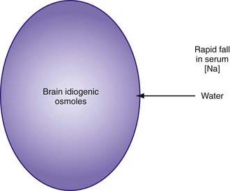
As hypernatremia develops, the brain generates idiogenic osmoles to increase the intracellular osmolality and prevent the loss of brain water. This mechanism is not instantaneous and is most prominent when hypernatremia has developed gradually. If the serum sodium concentration is lowered rapidly, there is movement of water from the serum into the brain cells to equalize the osmolality in the 2 compartments (Fig. 52-1). The resultant brain swelling manifests as seizures or coma.


Because of the associated dangers, hypernatremia should not be corrected rapidly. The goal is to decrease the serum sodium by <12 mEq/L every 24 hr, a rate of 0.5 mEq/L/hr. The most important component of correcting moderate or severe hypernatremia is frequent monitoring of the serum sodium value so that fluid therapy can be adjusted to provide adequate correction, neither too slow nor too fast. If a child has seizures as a result of brain edema secondary to rapid correction, administration of hypotonic fluid should be stopped. An infusion of 3% saline can acutely increase the serum sodium, reversing the cerebral edema.


In the child with hypernatremic dehydration, as in any child with dehydration, the first priority is restoration of intravascular volume with isotonic fluid (Chapter 54). Normal saline is preferable to lactated Ringer solution because the lower sodium concentration of the latter can cause the serum sodium to decrease too rapidly, especially if multiple fluid boluses are given. Repeated boluses of normal saline (10-20 mL/kg) may be required to treat hypotension, tachycardia, and signs of poor perfusion (peripheral pulses, capillary refill time) (Chapters 54 and 64).


The sodium concentration of the deficit replacement fluid, the rate of fluid administration, and the presence of continued water losses determine the rate of decrease of the sodium concentration. The following formula is often cited for calculating the water deficit:


image


This calculation is equivalent to 3-4 mL of water per kg for each 1 mEq that the current sodium level exceeds 145 mEq. The utility of such formulas has never been proven in clinical practice. Most patients with hypernatremic dehydration do well with a fluid sodium concentration of approximately half-normal saline, but with a fluid rate that is only 20-30% greater than maintenance fluid. Use of this concentration prevents excessive delivery of free water and too rapid a decrease in the serum sodium level. Patients with pure water loss may require a more hypotonic fluid (0.2 normal saline). Excessive water and sodium losses may also need to be replaced. If signs or symptoms of volume depletion develop, the patient receives additional boluses of isotonic saline. Monitoring of the rate of decrease of the serum sodium concentration permits adjustment in the rate and sodium concentration of the fluid that the patient is receiving, avoiding overly rapid correction of the hypernatremia. Many patients with mild to moderate hypernatremic dehydration due to gastroenteritis can be managed with oral rehydration (Chapter 332).


Acute, severe hypernatremia, usually secondary to sodium administration, can be corrected more rapidly because idiogenic osmoles have not had time to accumulate. This fact balances the high morbidity and mortality rates associated with hypernatremia with the dangers of overly rapid correction. When hypernatremia is severe and is due to sodium intoxication, it may be impossible to administer enough water to correct the hypernatremia rapidly without worsening the volume overload. In this situation, peritoneal dialysis allows for removal of the excess sodium. This requires dialysis fluid with a high glucose concentration and a low sodium concentration. In less severe cases, the addition of a loop diuretic increases the removal of excess sodium and water, decreasing the risk of volume overload. With sodium overload, hypernatremia is corrected with sodium-free intravenous fluid (5% dextrose in water [D5W]).


Hyperglycemia from hypernatremia is not usually a problem and is not treated with insulin because the acute decrease in glucose may precipitate cerebral edema by lowering plasma osmolality. Rarely, the glucose concentration of intravenous fluids must be reduced (from D5W to D2.5W). The secondary hypocalcemia is treated as needed.


It is important to address the underlying cause of the hypernatremia, if possible. The child with central diabetes insipidus should receive desmopressin acetate. Because this treatment reduces renal excretion of water, excessive intake of water must consequently be avoided to prevent both overly rapid correction of the hypernatremia and the development of hyponatremia. Over the long term, reduced sodium intake and the use of medications can somewhat ameliorate the water losses in nephrogenic diabetes insipidus (Chapter 524). The daily water intake of a child who is receiving tube feeding may need to be increased to compensate for high losses. The patient with significant ongoing losses, such as through diarrhea, may need supplemental water and electrolytes (Chapter 53). Sodium intake is reduced if it contributed to the hypernatremia.


Hyponatremia


Hyponatremia, a very common electrolyte abnormality in hospitalized patients, is a serum sodium level <135 mEq/L. Both total body sodium and TBW determine the serum sodium concentration. Hyponatremia exists when the ratio of water to sodium is increased. This condition can occur with low, normal, or high levels of body sodium. Similarly, body water can be low, normal, or high.


Etiology and Pathophysiology


The causes of hyponatremia are listed in Table 52-2. Pseudohyponatremia is a laboratory artifact that is present when the plasma contains very high concentrations of protein (multiple myeloma, intravenous immunoglobulin infusion) or lipid (hypertriglyceridemia, hypercholesterolemia). It does not occur when a direct ion-selective electrode determines the sodium concentration in undiluted plasma, a technique that is used by the instruments used for measuring arterial blood gases. In true hyponatremia, the measured osmolality is low, whereas it is normal in pseudohyponatremia. Hyperosmolality, as may occur with hyperglycemia, causes a low serum sodium concentration because water moves down its osmotic gradient from the intracellular space into the extracellular space, diluting the sodium concentration. However, because the manifestations of hyponatremia are due to the low plasma osmolality, patients with hyponatremia resulting from hyperosmolality do not have symptoms of hyponatremia. When the etiology of the hyperosmolality resolves, such as hyperglycemia in diabetes mellitus, water moves back into the cells and the sodium concentration rises to its “true” value. Mannitol or sucrose, a component of intravenous immunoglobulin preparations, may cause hyponatremia due to hyperosmolality.


Table 52-2 CAUSES OF HYPONATREMIA


PSEUDOHYPONATREMIA


EXTRARENAL LOSSES


RENAL LOSSES


EUVOLEMIC HYPONATREMIA


HYPERVOLEMIC HYPONATREMIA


MIM, database number from the Mendelian Inheritance in Man (http://www3.ncbi.nlm.nih.gov/Omim/).


* Most cases of proximal renal tubular acidosis are not due to this primary genetic disorder. Proximal renal tubular acidosis is usually part of Fanconi syndrome, which has multiple etiologies.


Classification of hyponatremia is based on the patient’s volume status. In hypovolemic hyponatremia, the child has lost sodium from the body. The water balance may be positive or negative, but sodium loss has been higher than water loss. The pathogenesis of the hyponatremia is usually a combination of sodium loss and water retention to compensate for the volume depletion. The patient has a pathologic increase in fluid loss, and this fluid contains sodium. Most fluid that is lost has a lower sodium concentration than that of plasma. Viral diarrhea fluid has, on average, a sodium concentration of 50 mEq/L. Replacing diarrhea fluid, which has a sodium concentration of 50 mEq/L, with formula, which has only approximately 10 mEq/L of sodium, reduces the sodium concentration. Intravascular volume depletion interferes with renal water excretion, the body’s usual mechanism for preventing hyponatremia. The volume depletion stimulates ADH synthesis, resulting in renal water retention. Volume depletion also decreases the GFR and enhances water resorption in the proximal tubule, thereby reducing water delivery to the collecting duct.


Diarrhea due to gastroenteritis is the most common cause of hypovolemic hyponatremia in children. Emesis causes hyponatremia if the patient takes in hypotonic fluid, either intravenously or enterally, despite the emesis. Most patients with emesis have either a normal sodium concentration or hypernatremia. Burns may cause massive losses of isotonic fluid and resultant volume depletion. Hyponatremia develops if the patient receives hypotonic fluid. Losses of sodium from sweat are especially high in children with cystic fibrosis, aldosterone deficiency, or pseudohypoaldosteronism, although high losses can occur simply in a hot climate. Third space losses are isotonic and can cause significant volume depletion, leading to ADH production and water retention, which can cause hyponatremia if the patient receives hypotonic fluid. In diseases that cause volume depletion through extrarenal sodium loss, the urine sodium level should be low (<10 mEq/L) as part of the renal response to maintain the intravascular volume. The only exceptions are diseases that cause both extrarenal and renal sodium losses: adrenal insufficiency and pseudohypoaldosteronism.


Renal sodium loss may occur in a variety of situations. In some situations, the urine sodium concentration is >140 mEq/L; thus, hyponatremia may occur without any fluid intake. In many cases, the urine sodium level is less than the serum concentration; thus, the intake of hypotonic fluid is necessary for hyponatremia to develop. In diseases associated with urinary sodium loss, the urine sodium level is >20 mEq/L despite volume depletion. This may not be true if the urinary sodium loss is no longer occurring, as is frequently the case if diuretics are discontinued. Because loop diuretics prevent generation of a maximally hypertonic renal medulla, the patient can neither maximally dilute nor concentrate the urine. The inability to maximally retain water provides some protection against severe hyponatremia. The patient receiving thiazide diuretics can concentrate the urine and is at higher risk for severe hyponatremia. Osmotic agents, such as glucose during diabetic ketoacidosis, cause loss of both water and sodium. Urea accumulates during renal failure and then acts as an osmotic diuretic after relief of urinary tract obstruction and during the polyuric phase of acute tubular necrosis. Transient tubular damage in these conditions further impairs sodium conservation. The serum sodium concentration in these conditions depends on the sodium concentration of the fluid used to replace the losses. Hyponatremia develops when the fluid is hypotonic relative to the urinary losses.


Renal salt wasting occurs in hereditary kidney diseases, such as juvenile nephronophthisis and autosomal recessive polycystic kidney disease. Obstructive uropathy, most commonly a consequence of posterior urethral valves, produces salt wasting, but patients with the disease may also have hypernatremia as a result of impaired ability to concentrate urine and high water loss. Acquired tubulointerstitial nephritis, usually secondary to either medications or infections, may cause salt wasting, along with other evidence of tubular dysfunction. CNS injury may produce cerebral salt wasting, which is theoretically due to the production of a natriuretic peptide that causes renal salt wasting. In type II renal tubular acidosis (RTA), usually associated with Fanconi syndrome (Chapter 523), there is increased excretion of sodium and bicarbonate in the urine. Patients with Fanconi syndrome also have glycosuria, aminoaciduria, and hypophosphatemia due to renal phosphate wasting.


Aldosterone is necessary for renal sodium retention and for the excretion of potassium and acid. In congenital adrenal hyperplasia due to 21-hydroxylase deficiency, the absence of aldosterone produces hyponatremia, hyperkalemia, and metabolic acidosis. In pseudohypoaldosteronism, aldosterone levels are elevated, but there is no response because of either a defective sodium channel or a lack of aldosterone receptors. A lack of tubular response to aldosterone may occur in children with urinary tract obstruction, especially during an acute urinary tract infection.


In hypervolemic hyponatremia, there is an excess of TBW and sodium, although the increase in water is greater than the increase in sodium. In most of the conditions that cause hypervolemic hyponatremia, there is a decrease in the effective blood volume, due to third space fluid loss, vasodilation, or poor cardiac output. The regulatory systems sense a decrease in effective blood volume and attempt to retain water and sodium to correct the problem. ADH causes renal water retention, and the kidney, under the influence of aldosterone and other intrarenal mechanisms, retains sodium. The patient’s sodium concentration decreases because water intake exceeds sodium intake and ADH prevents the normal loss of excess water.


In these disorders, there is a low urine sodium concentration (<10 mEq/L) and an excess of both TBW and sodium. The only exception is in patients with renal failure and hyponatremia. These patients have an expanded intravascular volume, and hyponatremia can therefore appropriately suppress ADH production. Water cannot be excreted because very little urine is being made. Serum sodium is diluted through ingestion of water. Because of renal dysfunction, the urine sodium concentration may be elevated, but urine volume is so low that urine sodium excretion has not kept up with sodium intake, leading to sodium overload. The urine sodium concentration in renal failure varies. In patients with acute glomerulonephritis, because it does not affect the tubules, the urine sodium level is usually low, whereas in patients with acute tubular necrosis, it is elevated because of tubular dysfunction.


Patients with hyponatremia and no evidence of volume overload or volume depletion have euvolemic hyponatremia. These patients typically have an excess of TBW and a slight decrease in total body sodium. Some of these patients have an increase in weight, implying that they are volume-overloaded. Nevertheless, from a clinical standpoint, they usually appear normal or have subtle signs of fluid overload.


In SIADH, the secretion of ADH is not inhibited by either low serum osmolality or expanded intravascular volume (Chapter 553). The result is that the child with SIADH is unable to excrete water. This results in dilution of the serum sodium and hyponatremia. The expansion of the extracellular volume due to the retained water causes a mild increase in intravascular volume. The kidney increases sodium excretion in an effort to decrease intravascular volume to normal; thus, the patients has a mild decrease in body sodium. SIADH most commonly occurs with disorders of the CNS (infection, hemorrhage, trauma, tumor, thrombosis), but lung disease (infection, asthma, positive pressure ventilation) and malignant tumors (producing ADH) are other potential causes. A variety of medications may cause SIADH, including recreational use of 3,4-methylenedioxymethylamphetamine (MDMA, or “Ecstasy”), opiates, antiepileptic drugs (carbamazepine, oxcarbamazepine, valproate), tricyclic antidepressants, vincristine, cytoxan, and selective serotonin reuptake inhibitors. The diagnosis of SIADH is one of exclusion, because other causes of hyponatremia must be eliminated (Table 52-3). Because SIADH is a state of intravascular volume expansion, low serum uric acid and BUN levels are supportive of the diagnosis.


A rare gain-of-function mutation in the renal ADH receptor causes nephrogenic syndrome of inappropriate antidiuresis. Patients with this X-linked disorder appear to have SIADH but have undetectable levels of ADH.


Hyponatremia in hospitalized patients is frequently due to inappropriate production of ADH and administration of hypotonic intravenous fluids. Causes of inappropriate ADH production include stress, medications such as narcotics or anesthetics, nausea, and respiratory illness. The synthetic analog of ADH, desmopressin acetate, causes water retention and may cause hyponatremia if fluid intake is not appropriately limited. The main uses of desmopressin acetate in children are for the management of central diabetes insipidus and of nocturnal enuresis.


Excess water ingestion can produce hyponatremia. In these cases, the sodium concentration decreases as a result of dilution. This decrease suppresses ADH secretion, and there is a marked water diuresis by the kidney. Hyponatremia develops only because the intake of water exceeds the kidney’s ability to eliminate water. This condition is more likely to occur in infants because their lower GFR limits their ability to excrete water. In some situations, the water intoxication causes acute hyponatremia and is due to a massive acute water load. Examples of causes of this water load include infant swimming lessons, inappropriate use of hypotonic intravenous fluids, water enemas, and forced water intake as a form of child abuse. Chronic hyponatremia occurs in children who receive water, but limited sodium and protein. The minimum urine osmolality is approximately 50 mOsm/kg, so the kidney can excrete 1 L of water only if there is enough solute ingested to produce 50 mOsm for urinary excretion. Because sodium and urea (a breakdown product of protein) are the principal urinary solutes, a lack of intake of sodium and protein prevents adequate water excretion. This occurs with the use of diluted formula or other inappropriate diets. Subsistence on beer, a poor source of sodium and protein, causes hyponatremia due to the inability to excrete the high water load (“beer potomania”).


The pathogenesis of the hyponatremia in glucocorticoid deficiency or hypothyroidism is incompletely understood. There is an inappropriate retention of water by the kidney, but the precise mechanisms are not clearly elucidated.


Clinical Manifestations


Hyponatremia causes a decrease in the osmolality of the extracellular space. Because the intracellular space then has a higher osmolality, water moves from the extracellular space to the intracellular space to maintain osmotic equilibrium. The increase in intracellular water causes cells to swell. Although cell swelling is not problematic in most tissues, it is dangerous for the brain, which is confined by the skull. As brain cells swell, there is an increase in intracranial pressure, which impairs cerebral blood flow. Acute, severe hyponatremia can cause brainstem herniation and apnea; respiratory support is often necessary. Brain cell swelling is responsible for most of the symptoms of hyponatremia. Neurologic symptoms of hyponatremia include anorexia, nausea, emesis, malaise, lethargy, confusion, agitation, headache, seizures, coma, and decreased reflexes. Patients may have hypothermia and Cheyne-Stokes respirations. Hyponatremia can cause muscle cramps and weakness; rhabdomyolysis can occur with water intoxication.


The symptoms of hyponatremia are mostly due to the decrease in extracellular osmolality and the resulting movement of water down its osmotic gradient into the intracellular space. Brain swelling can be significantly obviated if the hyponatremia develops gradually, because brain cells adapt to the decreased extracellular osmolality by reducing intracellular osmolality. This reduction is achieved by extrusion of the main intracellular ions (potassium and chloride) and a variety of small organic molecules. This process explains why the range of symptoms in hyponatremia is related to both the serum sodium level and its rate of decrease. A patient with chronic hyponatremia may have only subtle neurologic abnormalities with a serum sodium level of 110 mEq/L, but another patient may have seizures because of an acute decline in serum sodium level from 140 to 125 mEq/L.


Diagnosis


The history usually points to a likely etiology of the hyponatremia. Most patients with hyponatremia have a history of volume depletion. Diarrhea and diuretic use are very common causes of hyponatremia in children. A history of polyuria, perhaps with enuresis, and/or salt craving is present in children with primary kidney diseases or absence of aldosterone effect. Children may have signs or symptoms suggesting a diagnosis of hypothyroidism or adrenal insufficiency (Chapters 559 and 569). Brain injury raises the possibility of SIADH or cerebral salt wasting, with the caveat that SIADH is much more likely. Liver disease, nephrotic syndrome, renal failure, or congestive heart failure may be acute or chronic. The history should include a review of the patient’s intake, both intravenous and enteral, with careful attention to the amounts of water, sodium, and protein.


The traditional 1st step in the diagnostic process is determination of the plasma osmolality. This is done because some patients with a low serum sodium value do not have low osmolality. The clinical effects of hyponatremia are secondary to the associated low osmolality. Without a low osmolality, there is no movement of water into the intracellular space.


A patient with hyponatremia can have a low, normal, or high osmolality. A normal osmolality in combination with hyponatremia occurs in pseudohyponatremia. Children with elevation of serum glucose concentration or of another effective osmole (mannitol) have a high plasma osmolality and hyponatremia. The presence of a low osmolality indicates “true” hyponatremia. Patients with low osmolality are at risk for neurologic symptoms and require further evaluation to determine the etiology of the hyponatremia.


In some situations, true hyponatremia is present despite a normal or elevated plasma osmolality. The presence of an ineffective osmole, most commonly urea, increases the plasma osmolality, but because the osmole has the same concentration in the intracellular space, it does not cause fluid to move into the extracellular space. There is no dilution of the serum sodium by water, and the sodium concentration remains unchanged if the ineffective osmole is eliminated. Most importantly, the ineffective osmole does not protect the brain from edema due to hyponatremia. Hence, a patient may have symptoms of hyponatremia despite having a normal or increased osmolality because of uremia.


In patients with true hyponatremia, the next step in the diagnostic process is to clinically evaluate the volume status. Patients with hyponatremia can be hypovolemic, hypervolemic, or euvolemic. The diagnosis of volume depletion relies on the usual findings with dehydration (Chapter 54), although subtle volume depletion may not be clinically apparent. In a patient with subtle volume depletion, a fluid bolus results in a decrease in the urine osmolality and an increase in the serum sodium concentration. Children with hypervolemia are edematous on physical examination. They may have ascites, pulmonary edema, pleural effusion, or hypertension.


Hypovolemic hyponatremia can have renal or nonrenal causes. The urine sodium concentration is very useful in differentiating between renal and nonrenal causes. When the losses are nonrenal and the kidney is working properly, there is renal retention of sodium, a normal homeostatic response to volume depletion. Thus, the urinary sodium concentration is low, typically <10 mEq/L, although sodium conservation in neonates is less avid. When the kidney is the cause of the sodium loss, the urine sodium concentration is >20 mEq/L, reflecting the defect in renal sodium retention. The interpretation of the urine sodium level is challenging with diuretic therapy because it is high when diuretics are being used but low after the diuretic effect is gone. This becomes an issue only when diuretic use is surreptitious. The urine sodium concentration is not useful if a metabolic alkalosis is present; the urine chloride concentration must be used instead (Chapter 52.7).


Differentiating among the nonrenal causes of hypovolemic hyponatremia is usually facilitated by the history. Although the renal causes are more challenging to distinguish, a high serum potassium concentration is associated with disorders in which the sodium wasting is due to absence of or ineffective aldosterone.


In the patient with hypervolemic hyponatremia, the urine sodium concentration is a helpful parameter. It is usually <10 mEq/L, except in the patient with renal failure.


Treatment


The management of hyponatremia is based on the pathophysiology of the specific etiology. The management of all causes requires judicious monitoring and avoidance of an overly quick normalization of the serum sodium concentration. A patient with severe symptoms (seizures), no matter the etiology, should be given a bolus of hypertonic saline to produce a small, rapid increase in serum sodium. Hypoxia worsens cerebral edema, and hyponatremia may cause hypoxia. Hence, pulse oximetry should be monitored, and hypoxia aggressively corrected.


With all causes of hyponatremia, it is important to avoid “overly rapid” correction. The reason is that rapid correction of hyponatremia may cause central pontine myelinolysis (CPM). This syndrome, which occurs within several days of rapid correction of hyponatremia, produces neurologic symptoms, including confusion, agitation, flaccid or spastic quadriparesis, and death. There are usually characteristic pathologic and radiologic changes in the brain, especially in the pons.


CPM is more common in patients who are treated for chronic hyponatremia than in those treated for acute hyponatremia. Presumably, this difference is based on the adaptation of brain cells to the hyponatremia. The reduced intracellular osmolality that is an adaptive mechanism for chronic hyponatremia makes brain cells susceptible to dehydration during rapid correction of the hyponatremia, and this may be the mechanism of CPM. Even though CPM is rare in pediatric patients, it is advisable to avoid correcting the serum sodium concentration by >12 mEq/L/24 hr or > 18 mEq/L/48 hr. Desmopressin is a potential option if the serum sodium level is increasing too rapidly. This guideline does not apply to acute hyponatremia, as may occur with water intoxication, because the hyponatremia is more often symptomatic and there has not been time for the adaptive decrease in brain osmolality to occur. The consequences of brain edema in acute hyponatremia exceed the small risk of CPM.


Patients with hyponatremia can have severe neurologic symptoms, such as seizures and coma. The seizures associated with hyponatremia generally are poorly responsive to anticonvulsants. The child with hyponatremia and severe symptoms needs to receive treatment that will quickly reduce cerebral edema. This goal is best accomplished by increasing the extracellular osmolality so that water moves down its osmolar gradient from the intracellular space to the extracellular space.


Intravenous hypertonic saline rapidly increases serum sodium, and the effect on serum osmolality leads to a decrease in brain edema. Each mL/kg of 3% sodium chloride increases the serum sodium by approximately 1 mEq/L. A child with active symptoms often improves after receiving 4-6 mL/kg of 3% sodium chloride.


The child with hypovolemic hyponatremia has a deficiency in sodium and may have a deficiency in water. The cornerstone of therapy is to replace the sodium deficit and any water deficit that is present. The 1st step in treating any dehydrated patient is to restore the intravascular volume with isotonic saline. Ultimately, complete restoration of intravascular volume suppresses ADH production, thereby permitting excretion of the excess water. Chapter 54 discusses the management of hyponatremic dehydration.


The management of hypervolemic hyponatremia is difficult. Patients with this disorder have an excess of both water and sodium. Administration of sodium leads to worsening volume overload and edema. In addition, the patients are retaining water and sodium because of their ineffective intravascular volume or renal insufficiency. The cornerstone of therapy is water and sodium restriction, because the patients have volume overload. Diuretics may help by causing excretion of both sodium and water. Vasopressin antagonists (tolvaptan), by blocking the action of ADH and causing a water diuresis, are effective in correcting the hypervolemic hyponatremia due to heart failure or cirrhosis.


Some patients with low albumin due to nephrotic syndrome have a better response to diuretics after an infusion of 25% albumin; the sodium concentration often normalizes as a result of expansion of the intravascular volume. A child with congestive heart failure may have an increase in renal water and sodium excretion if there is an improvement in cardiac output. This improvement will “turn off” the regulatory hormones that are causing renal water (ADH) and sodium (aldosterone) retention. The patient with renal failure cannot respond to any of these therapies except fluid restriction. Insensible fluid losses eventually result in an increase in the sodium concentration as long as insensible and urinary losses are greater than intake. A more definitive approach in children with renal failure is to perform dialysis, which removes water and sodium.


In isovolumic hyponatremia, there is usually an excess of water and a mild sodium deficit. Therapy is directed at eliminating the excess water. The child with acute excessive water intake loses water in the urine because ADH production is turned off as a result of the low plasma osmolality. Children may correct their hyponatremia spontaneously over 3-6 hr. For acute, symptomatic hyponatremia due to water intoxication, hypertonic saline may be needed to reverse cerebral edema. For chronic hyponatremia due to poor solute intake, the child needs to receive an appropriate formula, and excess water intake should be eliminated.


Children with iatrogenic hyponatremia due to the administration of hypotonic intravenous fluids should receive 3% saline if they are symptomatic. Subsequent management is dictated by the patient’s volume status. The hypovolemic child should receive isotonic intravenous fluids. The child with nonphysiologic stimuli for ADH production should undergo fluid restriction. Prevention of this iatrogenic complication requires judicious use of intravenous fluids (Chapter 53).


Specific hormone replacement is the cornerstone of therapy for the hyponatremia of hypothyroidism or cortisol deficiency. Correction of the underlying defect permits appropriate elimination of the excess water.


SIADH is a condition of excess water, with limited ability of the kidney to excrete water. The mainstay of its therapy is fluid restriction. Furosemide is effective in the patient with SIADH and severe hyponatremia. Even in a patient with SIADH, furosemide causes an increase in water and sodium excretion. The loss of sodium is somewhat counterproductive, but this sodium can be replaced with hypertonic saline. Because the patient has a net loss of water and the urinary losses of sodium have been replaced, there is an increase in the sodium concentration, but no significant increase in blood pressure. Vasopressin antagonists (conivaptan, tolvaptan), which block the action of ADH and cause a water diuresis, are effective at correcting euvolemic hyponatremia, but overly rapid correction is a potential complication.


Treatment of chronic SIADH is challenging. Fluid restriction in children is difficult for nutritional and behavioral reasons. Other options are long-term furosemide therapy with sodium supplementation, an oral vasopressin antagonist (tolvaptan), or oral urea.


Bibliography


Centers for Disease Control and Prevention (CDC). Application of lower sodium intake recommendations to adults—United States, 1999-2006. MMWR Morb Mortal Wkly Rep. 2009;58:281-283.


Decaux G, Soupart A, Vassart G. Non-peptide arginine-vasopressin antagonists: the vaptans. Lancet. 2008;371:1624-1632.


Don M, Valerio G, Korppi M, et al. Hyponatremia in pediatric community-acquired pneumonia. Pediatr Nephrol. 2008;23:2247-2253.


Ellison DH, Berl T. The syndrome of inappropriate antidiuresis. N Engl J Med. 2007;356:2064-2072.


Fenske W, Maier SKG, Blechschmidt A, et al. Utility and limitations of the traditional diagnostic approach to hyponatremia: a diagnostic study. Am J Med. 2010;123:652-657.


Karakas HM, Erdem G, Yakinci C. Osmotic demyelination syndrome in a 40-day-old infant. Diagn Interv Radiol. 2007;13:121-124.


Leung C, Chang WC, Yeh SJ. Hypernatremic dehydration due to concentrated infant formula: report of two cases. Pediatr Neonatol. 2009;50:70-73.


Linshaw MA. Back to basics: congenital nephrogenic diabetes insipidus. Pediatr Rev. 2007;28:372-380.


Modi N. Avoiding hypernatraemic dehydration in healthy term infants. Arch Child. 2007;92:474-475.


Rianthavorn P, Cain JP, Turman MA. Use of conivaptan to allow aggressive hydration to prevent tumor lysis syndrome in a pediatric patient with large-cell lymphoma and SIADH. Pediatr Nephrol. 2008;23:1367-1370.


Rivkees SA. Differentiating appropriate antidiuretic hormone secretion, inappropriate antidiuretic hormone secretion and cerebral salt wasting: the common, uncommon, and misnamed. Curr Opin Pediatr. 2008;20:448-452.


Verbalis JG, Goldsmith SR, Greenberg A, et al. Hyponatremia treatment guidelines 2007: expert panel recommendations. Am J Med. 2007;120:S1-S21.


52.4 Potassium


Potassium Metabolism


Body Content and Physiologic Function


The intracellular concentration of potassium, approximately 150image mEq/L, is much higher than the plasma concentration (see Web Fig. 52-3). The majority of body potassium is contained in muscle. As muscle mass increases, there is an increase in body potassium. There is thus an increase in body potassium during puberty, and it is more significant in males. The majority of extracellular potassium is in bone; <1% of total body potassium is in plasma.


Because most potassium is intracellular, the plasma concentration does not always reflect the total body potassium content. A variety of conditions alter the distribution of potassium between the intracellular and extracellular compartments. The Na+,K+-ATPase maintains the high intracellular potassium concentration by pumping sodium out of the cell and potassium into the cell. This activity balances the normal leak of potassium out of cells via potassium channels that is driven by the favorable chemical gradient. Insulin increases potassium movement into cells by activating the Na+,K+-ATPase. Hyperkalemia stimulates insulin secretion, which helps mitigate the hyperkalemia. Acid-base status affects potassium distribution, probably via potassium channels and the Na+,K+-ATPase. A decrease in pH drives potassium extracellularly; an increase in pH has the opposite effect. β-Adrenergic agonists stimulate the Na+,K+-ATPase, increasing cellular uptake of potassium. This increase is protective, in that hyperkalemia stimulates adrenal release of catecholamines. α-Adrenergic agonists and exercise cause a net movement of potassium out of the intracellular space. An increase in plasma osmolality, as with mannitol infusion, leads to water movement out of the cells, and potassium follows as a result of solvent drag. The serum potassium concentration increases by approximately 0.6 mEq/L with each 10-mOsm rise in plasma osmolality.


The high intracellular concentration of potassium, the principal intracellular cation, is maintained via the Na+,K+-ATPase. The resulting chemical gradient is used to produce the resting membrane potential of cells. Potassium is necessary for the electrical responsiveness of nerve and muscle cells and for the contractility of cardiac, skeletal, and smooth muscle. The changes in membrane polarization that occur during muscle contraction or nerve conduction make these cells susceptible to changes in serum potassium levels. The ratio of intracellular to extracellular potassium determines the threshold for a cell to generate an action potential and the rate of cellular repolarization. The intracellular potassium concentration affects cellular enzymes. Potassium is necessary for maintaining cell volume because of its important contribution to intracellular osmolality.


Excretion


There is some loss of potassium in sweat, but it is normally minimal. The colon has the ability to eliminate some potassium. In addition, after an acute potassium load, much of the potassium, >40%, moves intracellularly, through the actions of epinephrine and insulin, which are produced in response to hyperkalemia. This process provides transient protection from hyperkalemia, but most ingested potassium is eventually excreted in the urine. The kidneys principally regulate long-term potassium balance, and they alter excretion in response to a variety of signals. Potassium is freely filtered at the glomerulus, but 90% is resorbed before the distal tubule and collecting duct, the principal sites of potassium regulation. The distal tubule and the collecting duct have the ability to absorb and secrete potassium. It is the amount of tubular secretion that regulates the amount of potassium that appears in the urine. The plasma potassium concentration directly influences secretion in the distal nephron. As the potassium concentration increases, secretion increases.


The principal hormone regulating potassium secretion is aldosterone, which is released by the adrenal cortex in response to increased plasma potassium. Its main site of action is the cortical collecting duct, where aldosterone stimulates sodium movement from the tubule into the cells. This movement creates a negative charge in the tubular lumen, facilitating potassium excretion. In addition, the increased intracellular sodium stimulates the basolateral Na+,K+-ATPase, causing more potassium to move into the cells lining the cortical collecting duct. Glucocorticoids, ADH, a high urinary flow rate, and high sodium delivery to the distal nephron also increase urinary potassium excretion. Potassium excretion is decreased by insulin, catecholamines, and urinary ammonia. Whereas ADH increases potassium secretion, it also causes water resorption, decreasing urinary flow. The net effect is that ADH has little overall impact on potassium balance. Alkalosis causes potassium to move into cells, including the cells lining the collecting duct. This movement increases potassium secretion, and because acidosis has the opposite effect, it decreases potassium secretion.


The kidney can dramatically vary potassium excretion in response to changes in intake. Normally, approximately 10-15% of the filtered load is excreted. In an adult, excretion of potassium can vary from 5-1,000 mEq/day.


Hyperkalemia


Hyperkalemia—because of the potential for lethal arrhythmias—is one of the most alarming electrolyte abnormalities.


Etiology and Pathophysiology


Three basic mechanisms cause hyperkalemia (Table 52-4). In the individual patient, the etiology is sometimes multifactorial.


Table 52-4 CAUSES OF HYPERKALEMIA


SPURIOUS LABORATORY VALUE


INCREASED INTAKE


TRANSCELLULAR SHIFTS


DECREASED EXCRETION


MIM, database number from the Mendelian Inheritance in Man (http://www3.ncbi.nlm.nih.gov/Omim/).


Fictitious or spurious hyperkalemia is very common in children because of the difficulties in obtaining blood specimens. This laboratory result is usually due to hemolysis during a heelstick or phlebotomy, but it can be the result of prolonged tourniquet application or fist clenching, either of which causes local potassium release from muscle.


The serum potassium level is normally 0.4 mEq/L higher than the plasma value, secondary to potassium release from cells during clot formation. This phenomenon is exaggerated with thrombocytosis because of potassium release from platelets. For every 100,000/m3 increase in the platelet count, the serum potassium level rises by approximately 0.15 mEq/L. This phenomenon also occurs with the marked white blood cell count elevations sometimes seen with leukemia. Elevated white blood cell counts, typically >200,000/m3, can cause a dramatic elevation in the serum potassium concentration. Analysis of a plasma sample usually provides an accurate result. It is important to analyze the sample promptly to avoid potassium release from cells, which occurs if the sample is stored in the cold, or cellular uptake of potassium and spurious hypokalemia, which occurs with storage of the sample at room temperature.


Because of the kidney’s ability to excrete potassium, it is unusual for excessive intake, by itself, to cause hyperkalemia. This condition can occur in a patient who is receiving large quantities of intravenous or oral potassium for excessive losses that are no longer present. Frequent or rapid blood transfusions can acutely increase the potassium level because of the potassium content of blood, which is variably elevated. Increased intake may precipitate hyperkalemia if there is an underlying defect in potassium excretion.


The intracellular space has a very high potassium concentration, so a shift of potassium from the intracellular space to the extracellular space can have a significant effect on the plasma potassium level. This shift occurs with metabolic acidosis, but the effect is minimal with an organic acid (lactic acidosis, ketoacidosis). A respiratory acidosis has less impact than a metabolic acidosis. Cell destruction, as seen with rhabdomyolysis, tumor lysis syndrome, tissue necrosis, or hemolysis, releases potassium into the extracellular milieu. The potassium released from red blood cells in internal bleeding, such as hematomas, is resorbed and enters the extracellular space.


Normal doses of succinylcholine or β-blockers and fluoride or digitalis intoxication all cause a shift of potassium out of the intracellular compartment. Succinylcholine should not be used during anesthesia in patients at risk for hyperkalemia. β-Blockers prevent the normal cellular uptake of potassium mediated by binding of β-agonists to the β2-adrenergic receptors. Potassium release from muscle cells occurs during exercise, and levels can increase by 1-2 mEq/L with high activity. With an increased plasma osmolality, water moves from the intracellular space and potassium follows. This process occurs with hyperglycemia, although in nondiabetic patients, the resultant increase in insulin causes potassium to move intracellularly. In diabetic ketoacidosis, the absence of insulin causes potassium to leave the intracellular space, and the problem is compounded by the hyperosmolality. The effect of hyperosmolality causes a transcellular shift of potassium into the extracellular space after mannitol or hypertonic saline infusions. Malignant hyperthermia, which is triggered by some inhaled anesthetics, causes muscle release of potassium (Chapter 603.2). Hyperkalemic periodic paralysis is an autosomal dominant disorder caused by a mutated sodium channel. It results in episodic cellular release of potassium and attacks of paralysis (Chapter 603.1).


The kidneys excrete most of the daily potassium intake, so a decrease in kidney function can cause hyperkalemia. Newborn infants in general, and especially premature infants, have decreased kidney function at birth and thus are at increased risk for hyperkalemia despite an absence of intrinsic renal disease. Neonates also have decreased expression of potassium channels, further limiting potassium excretion.


A wide range of primary adrenal disorders, both hereditary and acquired, can cause decreased production of aldosterone, with secondary hyperkalemia (Chapters 569 and 570). Patients with these disorders typically have metabolic acidosis and salt wasting with hyponatremia. Children with more subtle adrenal insufficiency may have electrolyte problems only during acute illnesses. The most common form of congenital adrenal hyperplasia, 21-hydroxylase deficiency, typically manifests in male infants as hyperkalemia, metabolic acidosis, hyponatremia, and volume depletion. Females with this disorder usually are diagnosed as newborns because of their ambiguous genitalia; treatment prevents the development of electrolyte problems.


Renin, via angiotensin II, stimulates aldosterone production. A deficiency in renin, a result of kidney damage, can lead to decreased aldosterone production. Hyporeninemia occurs in many kidney diseases, with some of the more common pediatric causes listed in Table 52-4. These patients typically have hyperkalemia and a metabolic acidosis, without hyponatremia. Some of these patients have impaired renal function, partially accounting for the hyperkalemia, but the impairment in potassium excretion is more extreme than expected for the degree of renal insufficiency.


A variety of renal tubular disorders impair renal excretion of potassium. Children with pseudohypoaldosteronism type 1 have hyperkalemia, metabolic acidosis, and salt wasting leading to hyponatremia and volume depletion; aldosterone values are elevated. In the autosomal recessive variant, there is a defect in the renal sodium channel that is normally activated by aldosterone. Patients with this variant have severe symptoms, beginning in infancy. Patients with the autosomal dominant form have a defect in the aldosterone receptor, and the disease is milder, often remitting in adulthood. Pseudohypoaldosteronism type 2, also called Gordon syndrome, is an autosomal dominant disorder characterized by hypertension due to salt retention and impaired excretion of potassium and acid, leading to hyperkalemia and metabolic acidosis. Activating mutations in either WNK1 or WNK4, both serine-threonine kinases located in the distal nephron, cause Gordon syndrome. In Bartter syndrome due to mutations in the potassium channel ROMK (type 2 Bartter syndrome), there can be transient hyperkalemia in neonates, but hypokalemia subsequently develops (Chapter 525).


Acquired renal tubular dysfunction, with an impaired ability to excrete potassium, occurs in a number of conditions. These disorders, all characterized by tubulointerstitial disease, are often associated with impaired acid secretion and a secondary metabolic acidosis. In some affected children, the metabolic acidosis is the dominant feature, although a high potassium intake may unmask the defect in potassium handling. The tubular dysfunction can cause renal salt wasting, potentially leading to hyponatremia. Because of the tubulointerstitial damage, these conditions may also cause hyperkalemia as a result of hyporeninemic hypoaldosteronism.

Stay updated, free articles. Join our Telegram channel

Jun 18, 2016 | Posted by in PEDIATRICS | Comments Off on Electrolyte and Acid-Base Disorders

Full access? Get Clinical Tree

Get Clinical Tree app for offline access