Fig. 13.1
Tubal ectopic pregnancy. An Inhomogeneous mass
)


A gestational sac with a yolk sac
A gestational sac with a yolk sac and foetal pole (Fig. 13.4

)
Fig. 13.4
Tubal ectopic pregnancy. A gestational sac with foetal pole
A gestational sac with a foetal pole and cardiac activity (Fig. 13.5

)
Fig. 13.5
Tubal ectopic pregnancy. A gestational sac with foetal pole and cardiac activity
The presence of foetal pole with cardiac activity outside the uterine cavity is highly specific for ectopic pregnancy. However the potential for false positive is higher with the presence of an empty sac or solid tubal mass [19, 66].
Other indirect ultrasound findings that could suggest ectopic pregnancy include:
- 1.
The presence of a pseudosac, which reflects the collection of fluid within the endometrial cavity. Small amount of bleeding within the uterine cavity is common in ectopic pregnancies, and it could resemble a small early intrauterine pregnancy. However, the pseudosac is usually surrounded with a single layer of endometrium unlike the gestational sac, which is surrounded by two layers or echogenic ring of trophoblast. As the pseudosac is the result of collection of fluid within the uterine cavity, it follows the contour of the cavity and tends to change in shape and moves position with scanning. This is different to true gestational sac that is implanted below the endometrial surface leaving an intact midline echo [59].
- 2.
The presence of an echogenic fluid in the pouch of Douglas. Echogenic fluid in the pelvis can suggest haemoperitoneum that can happen with ruptured ectopic pregnancy, but it can also relate to rupture haemorrhagic corpus luteum with an early intrauterine pregnancy [11
] (Fig. 13.6).

Fig. 13.6
Haemoperitoneum
13.5 Diagnosis of Non-tubal Ectopic Pregnancy
Despite the fact that the fallopian tube is the most common place for ectopic pregnancy, many other locations within the pelvis or abdomen have been identified. These are called non-tubal ectopic pregnancies and can happen in about 7 % of all ectopic pregnancies [17]. In recent years we have seen an increase in the prevalence of non-tubal pregnancies mainly due to increased number of Caesarean scar ectopic pregnancy, which is a consequence of the increasing rates of Caesarean section worldwide.
The significance of non-tubal ectopic pregnancies is that they are associated with higher morbidity and mortality rates when compared to tubal ectopic pregnancy. This is mainly due to the fact that they remain asymptomatic in the early weeks of gestations and present with major clinical symptoms in the late first or second trimester of pregnancy. In addition the symptoms and signs might not be typical of ectopic, and a correct ultrasound diagnosis is not always easy to achieve [40].
13.6 Interstitial Pregnancy
It occurs when the pregnancy implants in the interstitial part of the fallopian tubes. Although it is not common, approximately 2–6 % of all ectopics are interstitial [17], and the associated mortality rate is 2–2.5 %, which is much higher, compared to tubal ectopics [47].
The ultrasound features of interstitial pregnancies vary from a solid mass to the presence of a gestational sac with live embryo, but the pathognomonic diagnostic feature in ultrasound scan is the finding of the proximal part or interstitial part of the fallopian tube adjoining the lateral aspect of the uterine cavity to the medial aspect of the gestational sac. Interstitial pregnancy should not be confused with intrauterine pregnancies located high in the lateral aspect of the uterine cavity close to the tubal opening and with intrauterine pregnancies in women with congenital uterine anomalies. The identification of the interstitial parts of the fallopian tubes and the visible communication between the pregnancy and the uterine cavity can help to differentiate between these pregnancies [1, 29, 36].
The diagnosis can be further facilitated by the use of three-dimensional ultrasound, which provides superior images of the fundal aspect of the uterus and the interstitial section of the fallopian tubes [3

] (Figs. 13.7

and 13.8).
Fig. 13.7
Interstitial ectopic pregnancy
Fig. 13.8
3D Interstitial ectopic pregnancy
13.6.1 Cornual Pregnancy
Cornual pregnancy results from the implantation of the pregnancy in an atretic functional noncommunicating rudimentary cornu in women with unicornuate uteri. It is a rare form of ectopic pregnancy occurring in 1/76000 of all pregnancies [58].
Similar to interstitial pregnancy, the pregnancy can follow a silent presentation in the early weeks until uterine rupture and major internal bleeding occur, which typically occurs in the second trimester in about 50 % of cases. The typical sonographic features include an empty uterine cavity with a single interstitial tube. The gestational sac is separate from the main uterine corpus and surrounded by a thick wall representing the myometrium of the rudimentary cornu. A thick vascular pedicle connecting the empty corny and the gestational sac is another helpful sign [37, 51].
13.6.2 Abdominal Pregnancy
This occurs due to the implantation of the pregnancy in the peritoneal cavity outside the uterine cavity and the fallopian tube and ovaries. It happens in 1 % of all ectopic pregnancies and is related to high morbidity and mortality when compared to other types of ectopic pregnancy [7, 17].
Two theories behind this type of pregnancy have been described: the first type happens when the implantation of the pregnancy happens in the peritoneal cavity, and this is called a primary abdominal pregnancy, while a secondary abdominal pregnancy, which is more common, is the outcome of tubal miscarriage and reimplantation of the pregnancy in the abdominal cavity. Broad ligament with its high vascularity provides a good support for such type of pregnancy and thus is a common location for the implantation [7

, 17] (Fig. 13.9).
Fig. 13.9
Abdominal pregnancy
The ultrasound diagnosis includes empty uterine cavity and the identification of pregnancy sac separate from the pelvic organs. The ultrasound findings of a normal uterus with two interstitial portions of the tube help to differentiate between abdominal and cornual pregnancies. In advanced abdominal pregnancy, there is often severe foetal growth retardation and oligohydramnios [2, 8, 49].
13.6.3 Ovarian Pregnancy
This pregnancy is the result of pregnancy implanting in the ovarian substance or on the surface of the ovary and accounts for almost 3 % of all ectopic pregnancies [34

]. The clinical presentation is usually similar to tubal ectopic pregnancy, but the ultrasound morphology differs, and it includes the presence of a gestational sac surrounded by ovarian tissue and separate from the corpus luteum, which can be found in the ipsilateral or contralateral ovary. In addition there will be a negative sliding sign when trying to separate the gestational sac from the ovary on gentle palpation. A correct ultrasound diagnosis is not always possible especially when there is pelvic adhesions causing a tubal ectopic pregnancy to be adherent to the ovary and mimicking ovarian pregnancy [21, 61] (Fig. 13.10).
Fig. 13.10
Ovarian ectopic pregnancy
13.6.4 Cervical and Caesarean Scar Ectopic Pregnancy
Both of these ectopic pregnancies share the fact that they are the results of previous surgical trauma to the uterus. They are both characterised by trophoblast invasion beyond endometrial-myometrial junction, and they are classified as intramural pregnancies. Due to myometrial involvement, heavy bleeding often complicates surgical evacuation or even spontaneous expulsion [15].
Abnormal, painless vaginal bleeding is the main clinical presentation in these types of pregnancies. However, abdominal pain and even urinary symptoms caused by trophoblastic invasion into the bladder can occur in more advanced cases. Cervical pregnancy tends to be located low in the cervix in contrast to Caesarean scar pregnancy that is typically present close to the internal os [63, 73].
Correct ultrasound diagnosis is essential to differentiate between these types of ectopic pregnancies. It is even more important to differentiate them from a normal intrauterine pregnancy or inevitable miscarriage in order to avoid serious complications. The first ultrasound criterion for diagnosis is identifying the gestational sac or trophoblastic tissue at or below the level of the internal os with a clear invasion into the myometrium. Sliding organ sign will be negative as the gestational sac cannot be displaced from its surrounding. Another important ultrasound feature is the high blood flow (>20 cm/s) in the peritrophoblastic circulation on colour Doppler examination. This is different to inevitable miscarriage where the gestational sac usually changes position on applying gentle pressure with the ultrasound probe with no evidence of trophoblastic blood flow in the nearby myometrium [38, 60

,
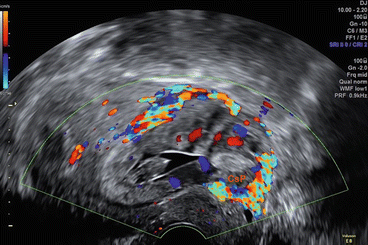
74


] (Figs. 13.11, 13.12, 13.13, 13.14 and 13.15).
Fig. 13.11
Early Caesarean scar ectopic pregnancy
Fig. 13.12
Caesarean scar ectopic pregnancy
Fig. 13.13
3D Caesarean scar ectopic pregnancy
Fig. 13.14
Cervical ectopic pregnancy
Fig. 10.15
3D Cervical ectopic pregnancy
The other differential diagnosis is low normal intrauterine pregnancy where the challenge will lie in differentiating the exact position of the pregnancy especially if it was very near to the internal os. This becomes even harder when the pregnancy is more advanced than the first trimester. However, the identification of the uterine artery insertion can be a useful guide to the level of the internal os, and the identification of myometrium invasion by trophoblastic tissue is usually the key factor in establishing the correct diagnosis [39, 72, 73].
13.7 Important Technical Tips
- 1
A transvaginal ultrasound scan is the best diagnostic test for women with suspected ectopic pregnancy. However, abdominal scans with empty bladder are helpful in women with large uteri due to fibroids or adenomyosis. Abdominal scans should also be considered in women with extensive adhesions between the uterus and anterior abdominal wall that typically occurs following multiple Caesarean sections. In these women uterine cavity is often not accessible on transvaginal scan.
- 2
Once a gestational sac is detected, it is important to make sure that it is intrauterine, and the best way to do it is to examine the uterus in longitudinal section to demonstrate continuity between the cervical canal and the location of the sac.
- 3
To distinguish intrauterine pregnancy from interstitial tubal pregnancy, check the interstitial parts of the fallopian tubes. If the gestational sac is located medially to the interstitial part of the tube, the pregnancy is intrauterine. In addition, a continuation between the endometrial cavity and the gestational sac is noted, which is not the case with interstitial pregnancy.
- 4
In women with enlarged uterus by fibroids that are distorting the shape of the uterine cavity, identifying the cervix first then following the cervical canal into the endometrial cavity can help to identify an intrauterine pregnancy.
- 5
In women with congenital uterine anomaly, to diagnose intrauterine and ectopic pregnancy, similar criteria that are used in women with normal uteri need to be applied. Bleeding can often occur in the empty part of the side of the anomalous uterine cavity, which could present itself as a pseudosac with a normal intrauterine pregnancy in the other side of the uterus.
- 6
In all cases a corpus luteum should be identified. This will help to locate ectopic pregnancy, as 80 % will be found on the ipsilateral side. In women with intrauterine pregnancy and more than one corpus luteum, a careful examination should be performed looking for a concomitant ectopic pregnancy.
- 7
Pouch of Douglas should be examined for the presence of blood. In most healthy pregnant women, there will be a small amount of clear or mildly echogenic fluid in the lesser pelvis. Blood clots appear solid and hyperechoic on ultrasound scan. They can be easily missed, as they appear similar to the echogenic bowel content. On palpation the clots have jelly-like consistency and there will be no signs of peristalsis. If severe intra-abdominal bleeding is suspected, a transabdominal examination of Morrison’s pouch (subhepatic space) should also be done.
13.8 Future Prospect
There is a considerable variation in the literature when it comes to the number of inconclusive scans in women presenting to early pregnancy units, with some units reporting the rate of inconclusive scans much higher than others. A published work by a group of experts has clearly identified that the prevalence of inconclusive ultrasound scans in different units has a strong relation to the quality of ultrasound examination. In the current situation where the standards of training among ultrasound users is very varied, raising the standards of early pregnancy scanning is needed to help improve the diagnosis of early pregnancy complications including ectopic pregnancy and thus improving management and reducing unnecessary interventions and also reduce the role of hCG in the diagnostic process.
Stay updated, free articles. Join our Telegram channel

Full access? Get Clinical Tree


