Chapter 24 Ectopic Pregnancy
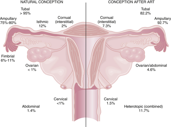
An ectopic pregnancy is a gestation that implants outside the endometrial cavity. Despite recent advances in earlier detection, it continues to represent a serious hazard to women’s health and their future reproductive potential. An ectopic pregnancy is estimated to occur in 1 of every 80 spontaneously conceived pregnancies. More than 95% of ectopic pregnancies implant in various anatomic segments of the fallopian tube, including the ampullary (75% to 80%), isthmic (12%), infundibular and fimbrial (6% to 11%), and interstitial (2%). Other, less common sites of ectopic implantation are the ovary, uterine cervix, and a rudimentary uterine horn. Rarely, an ectopic pregnancy may be intraligamentous or in the peritoneal cavity (abdominal pregnancy). With in vitro fertilization (IVF) and other assisted reproductive technologies (ARTs), the risk for ectopic pregnancy increases substantially, and the location of those ectopic implantations changes (Figure 24-1). Importantly, the risk for heterotropic implantations (one intrauterine and one ectopic) may rise to 1 in 100 with IVF. Other risk factors for an ectopic pregnancy include a history of a previous ectopic pregnancy, a pregnancy after tubal ligation or with an intrauterine device (IUD) in place, and a history of pelvic inflammatory disease (PID).

FIGURE 24-1 Possible locations of ectopic pregnancy with spontaneous conception vs pregnancies that result from assisted reproductive technologies (ART) such as in vitro fertilization (IVF).
(Modified from Pisarska MD and Carson SA: Incidence and risk factors for ectopic pregnancy. Clin Obstet Gynecol 42[1]:2-8,1999.)
Epidemiology and Etiology
Even though the mortality rates for ectopic pregnancy have dropped as a result of early diagnosis, this condition still causes 4% to 6% of maternal deaths in the United States and is the most common cause of maternal mortality in the first trimester.
In the past two decades, there was a significant increase in diagnosed ectopic pregnancy rates because of the following:
The key to the successful management of ectopic pregnancy is early diagnosis. A high index of suspicion and vigorous efforts at early diagnosis are needed, so “think ectopic!” is a sign that should be in every emergency room.
The etiology of ectopic pregnancy is not always clear but often is associated with known risk factors (Box 24-1). As many as half of cases result from an alteration of tubal transport mechanisms because of damage to the ciliated surface of the endosalpinx caused by infections, such as chlamydia and gonorrhea. Other etiologies include delayed fertilization, possible transmigration of the oocyte to the contralateral tube, and slowed tubal transport, which delays passage of the morula to the endometrial cavity. Chromosomal abnormalities of the fetus are not a cause of ectopic pregnancy.
Natural History of Untreated Tubal Ectopic Pregnancy
Tubal pregnancies rapidly invade the tubal mucosa, eroding into the tubal vessels, which are enlarged and engorged. The segment of the affected tube distends as the pregnancy grows and as blood from the eroded vessels dissects along the tubal wall. Women may have vaginal bleeding or spotting because the pregnancy hormones do not adequately support the endometrial lining and because bleeding from the tube may spill into the uterus as well as into the abdominal cavity. The most common outcomes of established tubal pregnancies include the following:
Symptoms and Clinical Diagnosis of Ectopic Tubal Pregnancy
The classic triad of symptoms of ectopic pregnancy consists of prior missed menses, vaginal bleeding, and lower abdominal pain. Clinical presentations represent a continuum: (1) acutely ruptured ectopic pregnancy, (2) probable ectopic pregnancy in a symptomatic woman, and (3) possible ectopic pregnancy. Each of these is discussed separately.
ACUTELY RUPTURED ECTOPIC PREGNANCY
The patient who has experienced rupture of her ectopic pregnancy most likely has intraperitoneal hemorrhage and presents with severe abdominal pain and dizziness. She may also complain of ipsilateral shoulder pain from phrenic nerve irritation caused by the blood in her upper abdomen. There may be signs of hemodynamic instability with tachycardia, diaphoresis, hypotension, and even loss of consciousness. Her abdomen may be distended and acutely tender, with guarding and rebound tenderness. The patient usually has cervical motion tenderness and a slightly enlarged, globular uterus. However, she may not have a palpable adnexal mass. The diagnosis is facilitated by a positive urine pregnancy test.
This clinical scenario represents a surgical emergency. Although other tests are often not necessary, an ultrasound would reveal an empty uterus and significant amounts of free fluid in the cul-de-sac. It is critical to establish large-bore intravenous lines and to start fluid resuscitation. Transfusion is important but should not delay emergency surgical intervention (usually by laparotomy, although laparoscopy may be appropriate when a patient is hemodynamically stable).
PROBABLE ECTOPIC PREGNANCY
Women who present with lower pelvic pain and vaginal spotting or bleeding, with or without amenorrhea, can be rapidly tested for pregnancy. The differential diagnosis includes threatened abortion or ectopic pregnancy. The patient generally has other clinical signs, such as tenderness of the abdomen with adnexal or cervical motion tenderness. The diagnosis of ectopic pregnancy may be confirmed by the absence of intrauterine pregnancy (IUP) on ultrasound in a woman with a level of human chorionic gonadotropin (hCG) that is sufficiently high to guarantee visualization of a normal IUP (see “Diagnostic Tests” later). There may be a variable amount of free fluid in the cul-de-sac detected by ultrasound. Only occasionally will the ectopic pregnancy be seen on ultrasound as a “double-ring sign” in the adnexa, but a corpus luteum cyst is often present. In such symptomatic women, even though they have stable vital signs, surgical exploration is generally recommended. Conservative surgical procedures, which preserve the fallopian tube, are generally indicated in women desiring future fertility (see “Management” later).
POSSIBLE ECTOPIC PREGNANCY
The most common clinical presentation is that of a possible ectopic pregnancy. Because her symptoms are so mild and nonspecific, a patient with an ectopic pregnancy may be seen at more than one visit before the diagnosis is confirmed.
Lower abdominal pain is present in most cases, although it may be mild. Amenorrhea or a history of an abnormal last menstrual period is obtained in 75% to 90% of ectopic pregnancies. Abnormal vaginal bleeding is seen in more than half of patients and ranges from spotting to the equivalent of a normal menstrual period. This spotting or bleeding results from an abnormally low production of hCG by the ectopic trophoblastic tissue. Distinguishing patients with ectopic pregnancy from those with an early threatened abortion or a spontaneous abortion can be challenging.
On physical examination, most patients are afebrile, and less than half have a discernable adnexal mass on pelvic examination. Often, the mass is palpated on the side opposite to the ectopic pregnancy and represents a corpus luteum in the contralateral ovary. The uterus is soft and is either of normal size or slightly enlarged. On ultrasound, there is a thickened endometrial stripe representing the visible sign of an Arias-Stella reaction, the histologic changes in the endometrial epithelium due to hCG stimulation. There may be a small amount of fluid seen in the cul-de-sac representing some slight intraperitoneal hemorrhage. Rarely is the ectopic pregnancy actually visualized.
DIFFERENTIAL DIAGNOSIS
Many gynecologic and nongynecologic disorders have symptoms in common with ectopic pregnancy and are listed in Box 24-2. A diagnostic algorithm for ectopic pregnancy is illustrated in Figure 24-2.

Stay updated, free articles. Join our Telegram channel

Full access? Get Clinical Tree

