CHAPTER 15 Duodenal Obstruction
Step 1: Surgical Anatomy
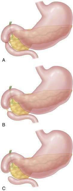
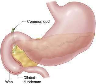
♦ In congenital duodenal atresia, the obstruction is distal to the ampulla of Vater in 85% of patients. A complete obstruction may be a luminal web (most common), an atresia connected by a fibrous cord, or complete separation of proximal and distal segments (Fig. 15-1).
♦ The ampulla is located close to the medial wall of the web, and care must be taken to avoid injuring it during repair.
♦ A fenestrated duodenal web with incomplete obstruction may not be noted at birth but can be seen later with feeding intolerance or obstruction when solid foods are started.
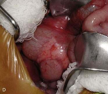
♦ Annular pancreas may accompany duodenal atresia. The annular portion of the pancreas can contain a significant pancreatic duct (Fig. 15-1, D).
Step 2: Preoperative Considerations
♦ The differential diagnosis includes malrotation with volvulus, small bowel atresia, and Hirschsprung disease in patients with bilious vomiting. In patients with nonbilious emesis, the differential includes pyloric atresia and antral web.
♦ Prenatal ultrasound and postnatal plain films show classic “double-bubble” configuration with a dilated first portion of the duodenum (Fig. 15-3).
Stay updated, free articles. Join our Telegram channel

Full access? Get Clinical Tree


