54 Disorders of Thrombosis and Hemostasis
The coagulation system is complex with intricately balanced interactions between the vascular endothelium, platelets, procoagulant, and anticoagulant proteins. With vascular injury, a cascade of interactions occurs between platelets and procoagulant proteins to initiate clot formation. After clot formation is initiated, anticoagulant proteins are activated to inhibit excessive clot formation. A dysregulation in this finely tuned system can lead to either a bleeding diathesis or a prothrombotic disorder.
Epidemiology
Congenital Bleeding Disorders
von Willebrand disease (vWD) is the most common congenital bleeding disorder, present in approximately 1% of the population. Hemophilia is the most common severe bleeding disorder and is the result of either a deficiency in factor VIII (hemophilia A) or IX (hemophilia B). The incidence of hemophilia is one in 5000 males, with 80% to 85% hemophilia A and 15% to 20% hemophilia B. There are no significant racial differences in the incidence of hemophilia.
Thrombosis
Advances in the treatment and support of critically ill children combined with an increasing awareness of thrombotic complications have likely resulted in an increase in the diagnosis of pediatric thromboembolic events. Although these events are increasing, they are still relatively rare compared with adults. This increase is most notable in comparing the rates from an older pediatric Canadian registry with an incidence of thromboembolic events of 5.3 in 10,000 hospitalizations, but a newer study from the United States reported a rate of 58 in 10,000 hospitalizations. There is a bimodal age distribution of thrombosis with peak rates in the neonatal and adolescent age groups.
Etiology And Pathogenesis
Congenital Bleeding Disorders
vWD is a family of disorders caused by quantitative or qualitative defects of von Willebrand factor (vWF), a plasma protein that plays a role in both platelet adhesion and fibrin formation.
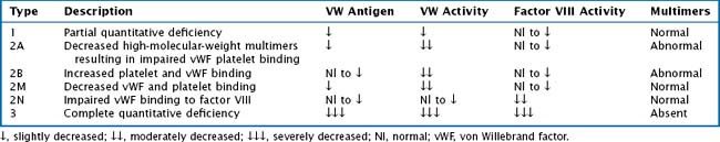
vWF is also a carrier protein for factor VIII; factor VIII unbound to vWF is rapidly degraded. vWD is classified into six types: 1, 2A, 2B, 2M, 2N, and 3. Table 54-1 provides a description and laboratory findings for all types of vWD.
Type 1 vWD is inherited in an autosomal dominant fashion and accounts for 70% to 80% of all vWD cases. It is characterized by a partial quantitative defect resulting in decreased amounts of a functionally normal vWF protein. Type 3 is a complete quantitative defect with no vWF and subsequently decreased factor VIII levels. The type 2 variants are qualitative defects.
Hemophilia A is a deficiency of factor VIII, and hemophilia B is a deficiency of factor IX. Both types of hemophilia are X-linked recessive diseases. About 30% of cases of hemophilia represent new mutations. Clinical severity is classified by the patient’s baseline factor level, severe less than 1%, moderate 1% to 5% and mild greater than 5%.
Acquired Bleeding Disorders
Children may develop bleeding disorders under various clinical conditions. The liver is responsible for the synthesis of most of the clotting factors. When synthetic liver dysfunction occurs, patients may develop abnormal coagulation profiles and clinical bleeding. Vitamin K plays a key role in the activation of coagulation proteins through the carboxylation of glutamate residues. Vitamin K deficiency can result from lack of exogenous vitamin K, as in an infant who is strictly breastfed or a child who is dependent on parenteral nutrition, or from infection or prolonged antibiotic administration. When vitamin K levels decline, the liver is unable to adequately synthesize functional factors II, VII, IX, and X, resulting in a bleeding propensity.
Disseminated Intravascular Coagulation
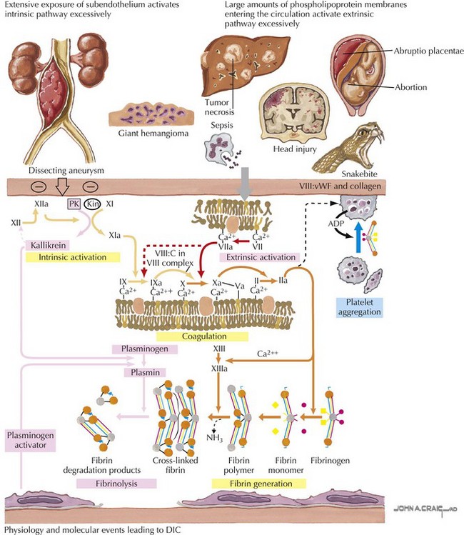
In disseminated intravascular coagulation (DIC), the normal physiology of coagulation is disturbed with resultant bleeding and thrombosis within the microvasculature. DIC is always secondary to a precipitating condition such as infection, tissue injury, malignancy, venom or toxin, or microangiopathic disorders. There is widespread deposition of intravascular fibrin leading to compromised organ function and capillary leak. Continued activation of this system leads to depletion of platelets and clotting factors, resulting in clinical bleeding (Figure 54-1).
Thrombosis
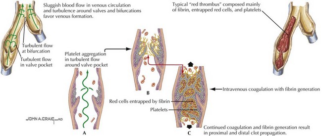
Deep venous thrombosis (DVT) is a consequence of Virchow’s triad of venous stasis, endothelial injury, and a hypercoagulable state. More than 90% of children with DVT have an identifiable prothrombotic risk factor. The most common risk factor is the presence of a central venous catheter (CVC). More than 50% of DVTs in children and more than 80% in neonates are associated with a CVC. Venous stasis in children may be caused by a postoperative state, casting or splinting, or other causes of prolonged immobility. Endothelial injury may be caused by trauma, infection, inflammation, or CVCs. Children may also have an inherited thrombophilia such as factor V Leiden or prothrombin gene mutations, deficiencies in the anticoagulant proteins including C, S, or antithrombin, hyperhomocysteinemia, or an elevated lipoprotein A. Acquired causes of thrombophilia include dehydration, antiphospholipid antibodies, malignancy, inflammatory states, anticoagulant deficiencies from consumption, or loss and exposure to certain medications such as estrogen-containing oral contraceptive pills or L-asparaginase (Figure 54-2).

Stay updated, free articles. Join our Telegram channel

Full access? Get Clinical Tree

