Disorders of the Neck and Spine

24 Disorders of the Neck and Spine


Pediatric disorders of the neck and spine are generally rare. The majority of these conditions can be treated with rest, antiinflammatory medications, targeted physical therapy, and orthotics. Operative management is typically reserved for severe or refractory pathology and symptoms.


Congenital Muscular Torticollis


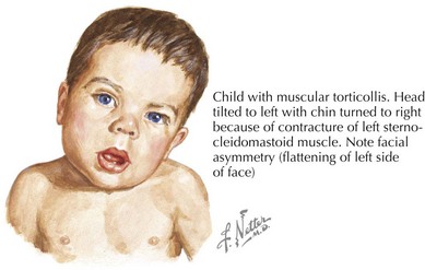
Torticollis, from the Latin words for “twisted neck,” is lateral neck flexion with contralateral head rotation (Figure 24-1). Although the majority of torticollis cases result from sternocleidomastoid muscle (SCM) shortening, other causes include neurologic conditions, cervical spine abnormalities, and vision or hearing deficits.


Congenital muscular torticollis (CMT) occurs in 0.3% to 2% of newborns. Although the cause is unknown, intrauterine crowding or birth trauma may cause muscle damage. More than half of infants with CMT are firstborn or had a history of difficult delivery, including use of forceps or breech presentation. Magnetic resonance imaging (MRI) and surgical histopathology have demonstrated muscle atrophy and fibrosis similar to the vascular complications of compartment syndrome. Abnormal head positioning in CMT can cause plagiocephaly and facial asymmetry.


Evaluation of CMT should include a thorough birth history and description of presenting symptoms. The physical examination classically demonstrates an infant with the head tilted towards the side of SCM contraction with the chin pointed in toward the opposite shoulder (see Figure 24-1). A soft, nontender mass may be palpable in the body of the SCM. Visual tracking, hearing, and neurologic function should also be assessed to rule out nonmuscular causes. Patients should be evaluated for other associated conditions such as developmental dysplasia of the hip and referred for diagnostic imaging or surgical consultation as necessary.


Cervical spine radiographs can rule out osseous causes, including congenital spinal fusion (e.g., Klippel-Feil syndrome, unilateral atlantooccipital fusion), hemivertebrae, and rotational cervical instability. Ultrasonography, the diagnostic modality of choice, demonstrates a hyperechogenic muscle mass. Computed tomography (CT) and MRI can help exclude suspected neurologic or osseous etiologies of torticollis. However, these modalities may not be as sensitive as ultrasonography in diagnosing CMT, and exposure to radiation or sedation limits their role in routine evaluation of torticollis.


The majority of CMT resolves spontaneously or with regular physical therapy. If muscle shortening persists beyond 6 months of therapy or 12 months of life, orthopedic referral is necessary for surgical SCM lengthening. Although successful CMT therapy typically resolves the associated plagiocephaly and facial asymmetry, patients with residual symptoms may require referral for further evaluation and treatment.


Scoliosis


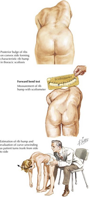
Scoliosis is a lateral curvature and rotation of the spine (Figure 24-2). Although most cases of scoliosis are idiopathic, it can also arise from congenital or neuromuscular disorders and is associated with various syndromes.


Idiopathic disease accounts for 80% of all scoliosis cases and is divided by age into infantile (0–3 years), juvenile (3–10 years), and adolescent (>10 years) groups. Infantile idiopathic scoliosis is rare and is unique in that it is more likely to occur in boys, have left-sided curvature, and spontaneously resolve. Juvenile idiopathic scoliosis is similar to the adolescent type; girls are predominantly affected, and the curvature is typically right-sided. Although juvenile idiopathic scoliosis does not occur during a period of rapid spine growth, it is more likely to be progressive than other types. Idiopathic scoliosis most commonly presents in adolescence (70%). Although the cause and pathogenesis of idiopathic scoliosis are largely unknown, there are multiple theories, including genetic factors; lack of melatonin; and abnormalities of platelets, nerves, and muscles.


The evaluation of idiopathic scoliosis should include a careful history and physical examination. Patients are screened for thoracolumbar asymmetry with an Adam’s forward bend test and scoliometer (see Figure 24-2). Examination should include comparison of the shoulders, scapulae, trunk, pelvis, and legs. Because idiopathic scoliosis is a diagnosis of exclusion, a careful assessment of neurologic function and signs of associated orthopedic disorders should be assessed. Congenital scoliosis should not be mistaken for infantile idiopathic scoliosis because the former is associated with other osseous and visceral anomalies. Definitive diagnosis and disease severity are determined by measuring the Cobb angle on a posteroanterior spinal radiograph, with any curvature more than 10 degrees being diagnostic. MRI is typically reserved for an atypical presentation or patients with focal neurologic findings.
Treatment of patients with idiopathic scoliosis depends on the age of onset and degree of curvature. Children with mild to moderate curves (<25 degrees), especially in the skeletally mature, can be observed clinically and with serial spinal radiographs. Physical therapy may also be used for minor disease. Patients with more severe curves, particularly younger children, may require bracing to minimize curve progression and prevent potential surgical correction. Unfortunately, the evidence for physical therapy and orthosis is equivocal, and their use is variable. Surgical correction is reserved for patients with severe curvatures (>45 degrees) or progressive disease that could compromise pulmonary or musculoskeletal function. Surgery can improve both the physical and psychological sequelae of idiopathic scoliosis. However, patients and their parents should be cautioned that although operative management can provide permanent structural correction, there are risks of surgical side effects or persistent symptoms.

< div class='tao-gold-member'>

Stay updated, free articles. Join our Telegram channel

Jun 19, 2016 | Posted by in PEDIATRICS | Comments Off on Disorders of the Neck and Spine

Full access? Get Clinical Tree

Get Clinical Tree app for offline access