103 Disorders of the Gastrointestinal System
Gastrointestinal (GI) disorders are common causes of serious illness in the neonatal population. Necrotizing enterocolitis (NEC) affects 1% to 8% of all infants in neonatal intensive care units (NICUs), and population-based reports not limited to the NICU indicate that anywhere from one in 700 to one in 10,000 infants will experience GI obstruction. Neonatal GI disorders share many common and nonspecific presenting signs. However, careful history regarding the timing of symptom onset, relation to initiation of feeds, and maternal and perinatal factors can direct an efficient diagnostic evaluation and initial management.
Necrotizing Enterocolitis
Etiology and Pathogenesis
Necrotizing enterocolitis (NEC) is one of the most common GI conditions affecting premature neonates. Despite advances made in the diagnosis and treatment of other conditions associated with prematurity, rates of NEC have increased in the past several decades because of the increased survival of infants at younger gestational ages. Mortality associated with NEC remains significant, ranging from 10% to 50%.
Although the pathogenesis of NEC is not clear, the concurrence of multiple conditions seems to result in NEC. Prematurity is the most consistently identified risk factor. The association of NEC with initiation of feeds, osmolarity of feeds, and rate of feed increases suggests that immature gut mucosa is injured in a way that allows intestinal bacterial translocation. This results in an immune response that may progress to a systemic inflammatory response and shock. Clusters of NEC cases in NICUs and the isolation of bacteria from infants affected by NEC suggests that microbial infection may play a role in either initiating or propagating the above cascade of events. Bacteria isolated from infants affected by NEC include Klebsiella pneumoniae, Pseudomonas aeruginosa, Escherichia coli, and Enterobacter spp.
Clinical Presentation
Early signs of NEC are nonspecific, and a high index of suspicion is required for early identification. Feeding intolerance, abdominal distension, lethargy, and temperature instability are early harbingers of evolving NEC. Later findings include bloody stools; worsening abdominal distension; abdominal skin redness and necrosis and; in severe cases, findings associated with viscus perforation and fulminant sepsis.
Evaluation and Management
Radiographic findings associated with NEC are variable. Early NEC can present with isolated bowel loop distension. The most classic sign of NEC seen on abdominal radiographs is pneumatosis intestinalis (i.e., intramural air). In the most severe cases, abdominal free air becomes evident. In cases in which microperforation results in release of air into the venous system, the only radiographic finding that may be detected is portal venous gas.
The most common laboratory finding is a low white blood cell count. However, thrombocytopenia, acidosis, and hypoglycemia are also seen. In cases that evolve to septic shock, laboratory study results may be consistent with disseminated intravascular coagulation.
The majority of infants affected by NEC are managed medically, the principles of which include supportive care followed by a period of bowel rest and slow reintroduction of feeds. Patients should be followed closely for progression of disease, which is evident from enlarging abdominal circumference, declining clinical status, and evolution of radiographic findings. Because physical findings may be subtle, repeated abdominal imaging is often performed in the early stages of NEC to identify evolving disease.
Intubation and ventilatory support may be required. Acidosis is a marker of progressive disease and inadequate perfusion of end organs. When needed, pressors should be initiated to maintain hemodynamic stability. After blood cultures are drawn, empiric antibiotic treatment for intestinal flora should be started. Although ampicillin, gentamicin, and Flagyl have been the first-line choice of treatment, local antibiotic susceptibilities should guide therapy. Infants should receive no enteral nutrition for a period of time, typically 7 to 14 days, after which feeds should be gradually introduced.
Approximately one-third to one-half of patients with NEC require surgical management. Indications for surgical management include intestinal perforation or bowel necrosis. The preferred approach in infants weighing more than 1500 g is resection of necrotic bowel and temporary diversion to a mucocutaneous fistula, with reanastomosis after approximately 6 weeks. In patients weighing less than1500 g, primary peritoneal drainage is used because of significant morbidity and mortality associated with resection and diversion.
Several complications are associated with NEC. The most common is intestinal stricture, which presents with feeding intolerance about 1 to 2 weeks after the resolution of NEC. This should be distinguished from recurrence of NEC, which is seen in about 6% of NEC survivors. Other long-term complications include neurodevelopmental delay as well as total parenteral nutrition–associated cholestasis in infants who are on prolonged parenteral nutrition during treatment for NEC. Importantly, if a lengthy section of intestine required resection, infants can experience short gut syndrome.
Intestinal Obstruction
Etiology and Pathogenesis
Intestinal obstruction is possible at any point along the GI tract. Causes of obstruction are subdivided into mechanical and functional causes. Mechanical causes result from a failure of development of the GI tract. Examples include intestinal and anal atresias, which are caused by a failure of recanalization of the alimentary tract; malrotation, which is caused by a failure of complete rotation of the GI tract during development; and pyloric stenosis, which is caused by overgrowth of the pyloric muscle after birth. Functional causes of obstruction include meconium plug syndrome, Hirschsprung’s disease, and small left colon syndrome, all of which result from impairment of the normal peristaltic movements.
Clinical Presentation
Intestinal obstruction in a neonate, regardless of the underlying cause, presents with the following common signs:
Subtle differences in presentation can suggest the location of the obstruction along the alimentary tract (Table 103-1). These unique features are highlighted below as each disorder is discussed.
Table 103-1 Localizing Features of Intestinal Obstruction
Sign or Finding | Proximal or Distal | Specific Site (When Applicable) |
---|---|---|
Nonbilious vomiting | Proximal | Proximal to the ampulla of Vater |
Bilious vomiting | Proximal or distal | Distal to the ampulla of Vater |
Scaphoid abdomen | Proximal | Often preduodenal |
Distended abdomen | Distal | |
Maternal polyhydramnios | Proximal |
Esophageal Atresia
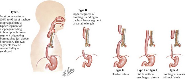
The esophagus can be obstructed in the form of esophageal atresia (EA), which is caused by a failure of separation of the esophagus from the trachea during normal development. Pure EA is rare and often coexists with tracheoesophageal fistula (TEF). A standard classification scheme describes the relationship between the atresia and the coexistent TEF (Figure 103-1). The most common combination is EA with distal TEF (type C). The most difficult to recognize and diagnose is “H-type,” which refers to isolated TEF without EA.

Stay updated, free articles. Join our Telegram channel

Full access? Get Clinical Tree

